46
views
0
recommends
+1 Recommend
1 collections
    0
    shares
      Are you tired of sifting through news that doesn't interest you?
      Personalize your Karger newsletter today and get only the news that matters to you!

      Sign up

      • Record: found
      • Abstract: found
      • Article: found
      Is Open Access

      Case of Unilateral Peripheral Cone Dysfunction

      case-report

      Read this article at

      ScienceOpenPublisherPMC
      Bookmark
          There is no author summary for this article yet. Authors can add summaries to their articles on ScienceOpen to make them more accessible to a non-specialist audience.

          Abstract

          Purpose

          Peripheral cone dystrophy is a subgroup of cone dystrophy, and only 4 cases have been reported. We present a patient with unilateral peripheral cone dysfunction and report the functional changes determined by electrophysiological tests and ultrastructural changes determined by spectral domain optical coherence tomography (SD-OCT).
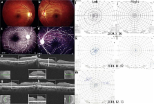
          Case

          A 34-year-old woman complained of blurred vision in both eyes. Our examination showed that her visual acuity was 0.05 OD and 0.2 OS. A relative afferent pupillary defect was present in her right eye. The results of slit-lamp examination, ophthalmoscopy, and fluorescein angiography were normal except for pallor of the right optic disc. SD-OCT showed a diffuse thinning of the retina in the posterior pole of the right eye. A severe constriction of the visual fields was found in both eyes but more in the right eye. The photopic full-field electroretinograms (ERGs) were reduced in the right eye but normal in the left eye. The multifocal ERGs were severely reduced throughout the visual field except in the central area of the right eye. The multifocal ERGs from the left eye were normal. The pattern visual evoked responses were within the normal range in both eyes. She had a 5-year history of sniffing paint thinner.

          Results

          Although the visual dysfunction was initially suspected to be due to psychological problems from the results of subjective tests, objective tests indicated a peripheral cone dysfunction in the right eye. The pathophysiological mechanism and the relationship with thinner sniffing were not determined.

          Conclusions

          Our findings indicate that peripheral cone dysfunction can occur unilaterally. Electrophysiology and SD-OCT are valuable tests to perform to determine the pathogenesis of unusual ocular findings objectively.

          Related collections

          Most cited references22

          • Record: found
          • Abstract: found
          • Article: not found

          Dominant mutations in RP1L1 are responsible for occult macular dystrophy.

          Occult macular dystrophy (OMD) is an inherited macular dystrophy characterized by progressive loss of macular function but normal ophthalmoscopic appearance. Typical OMD is characterized by a central cone dysfunction leading to a loss of vision despite normal ophthalmoscopic appearance, normal fluorescein angiography, and normal full-field electroretinogram (ERGs), but the amplitudes of the focal macular ERGs and multifocal ERGs are significantly reduced at the central retina. Linkage analysis of two OMD families was performed by the SNP High Throughput Linkage analysis system (SNP HiTLink), localizing the disease locus to chromosome 8p22-p23. Among the 128 genes in the linkage region, 22 genes were expressed in the retina, and four candidate genes were selected. No mutations were found in the first three candidate genes, methionine sulfoxide reductase A (MSRA), GATA binding 4 (GATA4), and pericentriolar material 1 (PCM1). However, amino acid substitution of p.Arg45Trp in retinitis pigmentosa 1-like 1 (RP1L1) was found in three OMD families and p.Trp960Arg in a remaining OMD family. These two mutations were detected in all affected individuals but in none of the 876 controls. Immunohistochemistry of RP1L1 in the retina section of cynomolgus monkey revealed expression in the rod and cone photoreceptor, supporting a role of RP1L1 in the photoreceptors that, when disrupted by mutation, leads to OMD. Identification of RP1L1 mutations as causative for OMD has potentially broader implications for understanding the differential cone photoreceptor functions in the fovea and the peripheral retina. 2010 The American Society of Human Genetics. Published by Elsevier Inc. All rights reserved.
            Bookmark
            • Record: found
            • Abstract: found
            • Article: not found

            Photoreceptor outer segment abnormalities as a cause of blind spot enlargement in acute zonal occult outer retinopathy-complex diseases.

            To investigate the correlation between visual field (VF) defects in diseases of the acute zonal occult outer retinopathy (AZOOR)-complex and their spectral-domain optical coherence tomographic (OCT) findings. Observational case series. Patients with AZOOR, multiple evanescent white dot syndrome (MEWDS), and multifocal choroiditis and panuveitis (MCP) examined in a private practice retinal referral center had threshold VF testing and spectral-domain OCT examination performed using a device capable of obtaining a block of 128 B-scans in a 6 x 6-mm region centered on the optic nerve and macula. The areas of defects in the boundary between the inner segments (IS) and the outer segments (OS) of the photoreceptors, termed the IS/OS boundary, were compared with the VF defects measured. There were 18 evaluable eyes among one patient with MEWDS, two with AZOOR, and seven with MCP. In the 14 eyes with blind spot enlargement [corrected] corresponding IS/OS boundary defects were found in the [corrected] peripapillary region, while no IS/OS boundary defects were found in the four [corrected] eyes without blind spot enlargement. IS/OS boundary defects were seen over chorioretinal scars and areas of neovascularization and no widespread defects were seen [corrected] elsewhere in the fundus. The IS/OS boundary defects showed improvement, as did the blind spot enlargement, spontaneously in the patient with MEWDS and after treatment with immunosuppression in the patients with AZOOR. The spectral-domain OCT finding of IS/OS boundary defects, implicating photoreceptor OS perturbation, appears to explain the blind spot enlargement in patients with AZOOR-complex diseases. These defects are not necessarily permanent.
              Bookmark
              • Record: found
              • Abstract: found
              • Article: not found

              Acute zonal occult outer retinopathy. Donders Lecture: The Netherlands Ophthalmological Society, Maastricht, Holland, June 19, 1992.

              J Gass (1993)
              This report describes 13 patients, predominantly young women, with a syndrome characterized by rapid loss of one or more large zones of outer retinal function, photopsia, minimal funduscopic changes, and electroretinographic abnormalities affecting one or both eyes. All patients on follow-up examination had persistent visual field defects, and most had chronic photopsia and zones of pigment epithelial atrophy. Evidence is presented that these patients probably represent part of the spectrum of a single disorder that includes the multiple evanescent white-dot syndrome (MEWDS), acute idiopathic blind-spot-enlargement syndrome (AIBSES), acute macular neuroretinopathy (AMN), and the pseudo-presumed ocular histoplasmosis syndrome (P-POHS). The medical records of these 13 patients and 2 additional patients, who developed, in addition to the features of this syndrome, funduscopic changes typical of MEWDS, AMN, and P-POHS, were reviewed and follow-up obtained. These patients had extensive unrewarding medical and neurological investigations because of suspected diagnoses, including central nervous system disorders, cancer-associated retinopathy, retinal vasculitis, diffuse unilateral subacute neuroretinitis, and tapetoretinal degenerations. Although most patients retained good visual acuity, all had permanent visual field loss that in some cases was severe. The cause of the disorder was not determined. No effective treatment was found. Acute visual loss and photopsia in these patients is probably caused by damage to large zones of the outer retina that appears unaffected ophthalmoscopically. Electroretinography is important in early diagnosis. Future investigations probably will reveal further evidence linking this disorder to MEWDS, AIBSES, AMN, and P-POHS.
                Bookmark

                Author and article information

                Journal
                Case Report Ophthalmol
                Case Report Ophthalmol
                COP
                Case Reports in Ophthalmology
                S. Karger AG (Allschwilerstrasse 10, P.O. Box · Postfach · Case postale, CH–4009, Basel, Switzerland · Schweiz · Suisse, Phone: +41 61 306 11 11, Fax: +41 61 306 12 34, karger@karger.ch )
                1663-2699
                May-Aug 2012
                10 May 2012
                10 May 2012
                : 3
                : 2
                : 162-168
                Affiliations
                [1] aDepartment of Ophthalmology, Juntendo University Urayasu Hospital, Chiba, Tokyo, Japan
                [2] bDepartment of Ophthalmology, Teikyo University School of Medicine, University Hospital Itabashi, and Tokyo, Japan
                [3] cCarl Zeiss Meditec Co., Ltd., Tokyo, Japan
                Author notes
                *Kei Shinoda, MD, Department of Ophthalmology, Teikyo University School of Medicine, 2-11-1 Kaga, Itabashi-ku, Tokyo 173-8605 (Japan), Tel. +81 3 3964 1225, E-Mail shinodak@ 123456med.teikyo-u.ac.jp
                Article
                cop-0003-0162
                10.1159/000339129
                3369245
                22679434
                e4240684-7c00-4507-aedd-bb16b898554e
                Copyright © 2012 by S. Karger AG, Basel

                This is an Open Access article distributed under the terms of the Creative Commons Attribution-Noncommercial-No-Derivative-Works License ( http://creativecommons.org/licenses/by-nc-nd/3.0/). Users may download, print and share this work on the Internet for noncommercial purposes only, provided the original work is properly cited, and a link to the original work on http://www.karger.com and the terms of this license are included in any shared versions.

                History
                Page count
                Figures: 2, References: 22, Pages: 7
                Categories
                Published online: May, 2012

                Ophthalmology & Optometry
                tunnel vision,optical coherence tomography,multifocal erg,electroretinogram,pattern visual evoked potentials

                Comments

                Comment on this article