22
views
0
recommends
+1 Recommend
0 collections
    0
    shares
      • Record: found
      • Abstract: found
      • Article: found
      Is Open Access

      Omega-3 polyunsaturated fatty acid attenuates the inflammatory response by modulating microglia polarization through SIRT1-mediated deacetylation of the HMGB1/NF-κB pathway following experimental traumatic brain injury

      research-article

      Read this article at

      Bookmark
          There is no author summary for this article yet. Authors can add summaries to their articles on ScienceOpen to make them more accessible to a non-specialist audience.

          Abstract

          Background

          Microglial polarization and the subsequent neuroinflammatory response are contributing factors for traumatic brain injury (TBI)-induced secondary injury. High mobile group box 1 (HMGB1) mediates the activation of the NF-κB pathway, and it is considered to be pivotal in the late neuroinflammatory response. Activation of the HMGB1/NF-κB pathway is closely related to HMGB1 acetylation, which is regulated by the sirtuin (SIRT) family of proteins. Omega-3 polyunsaturated fatty acids (ω-3 PUFA) are known to have antioxidative and anti-inflammatory effects. We previously demonstrated that ω-3 PUFA inhibited TBI-induced microglial activation and the subsequent neuroinflammatory response by regulating the HMGB1/NF-κB signaling pathway. However, no studies have elucidated if ω-3 PUFA affects the HMGB1/NF-κB pathway in a HMGB1 deacetylation of dependent SIRT1 manner, thus regulating microglial polarization and the subsequent neuroinflammatory response.

          Methods

          The Feeney DM TBI model was adopted to induce brain injury in rats. Modified neurological severity scores, rotarod test, brain water content, and Nissl staining were employed to determine the neuroprotective effects of ω-3 PUFA supplementation. Assessment of microglia polarization and pro-inflammatory markers, such as tumor necrosis factor (TNF)-α, interleukin (IL)-1β, IL-6, and HMGB1, were used to evaluate the neuroinflammatory responses and the anti-inflammatory effects of ω-3 PUFA supplementation. Immunofluorescent staining and western blot analysis were used to detect HMGB1 nuclear translocation, secretion, and HMGB1/NF-κB signaling pathway activation to evaluate the effects of ω-3 PUFA supplementation. The impact of SIRT1 deacetylase activity on HMGB1 acetylation and the interaction between HMGB1 and SIRT1 were assessed to evaluate anti-inflammation effects of ω-3 PUFAs, and also, whether these effects were dependent on a SIRT1-HMGB1/NF-κB axis to gain further insight into the mechanisms underlying the development of the neuroinflammatory response after TBI.

          Results

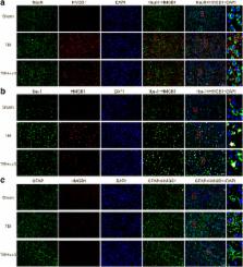
          The results of our study showed that ω-3 PUFA supplementation promoted a shift from the M1 microglial phenotype to the M2 microglial phenotype and inhibited microglial activation, thus reducing TBI-induced inflammatory factors. In addition, ω-3 PUFA-mediated downregulation of HMGB1 acetylation and its extracellular secretion was found to be likely due to increased SIRT1 activity. We also found that treatment with ω-3 PUFA inhibited HMGB1 acetylation and induced direct interactions between SIRT1 and HMGB1 by elevating SIRT1 activity following TBI. These events lead to inhibition of HMGB1 nucleocytoplasmic translocation/extracellular secretion and alleviated HMGB1-mediated activation of the NF-κB pathway following TBI-induced microglial activation, thus inhibiting the subsequent inflammatory response.

          Conclusions

          The results of this study suggest that ω-3 PUFA supplementation attenuates the inflammatory response by modulating microglial polarization through SIRT1-mediated deacetylation of the HMGB1/NF-κB pathway, leading to neuroprotective effects following experimental traumatic brain injury.

          Related collections

          Most cited references39

          • Record: found
          • Abstract: found
          • Article: not found

          HDAC inhibition prevents white matter injury by modulating microglia/macrophage polarization through the GSK3β/PTEN/Akt axis.

          Severe traumatic brain injury (TBI) elicits destruction of both gray and white matter, which is exacerbated by secondary proinflammatory responses. Although white matter injury (WMI) is strongly correlated with poor neurological status, the maintenance of white matter integrity is poorly understood, and no current therapies protect both gray and white matter. One candidate approach that may fulfill this role is inhibition of class I/II histone deacetylases (HDACs). Here we demonstrate that the HDAC inhibitor Scriptaid protects white matter up to 35 d after TBI, as shown by reductions in abnormally dephosphorylated neurofilament protein, increases in myelin basic protein, anatomic preservation of myelinated axons, and improved nerve conduction. Furthermore, Scriptaid shifted microglia/macrophage polarization toward the protective M2 phenotype and mitigated inflammation. In primary cocultures of microglia and oligodendrocytes, Scriptaid increased expression of microglial glycogen synthase kinase 3 beta (GSK3β), which phosphorylated and inactivated phosphatase and tensin homologue (PTEN), thereby enhancing phosphatidylinositide 3-kinases (PI3K)/Akt signaling and polarizing microglia toward M2. The increase in GSK3β in microglia and their phenotypic switch to M2 was associated with increased preservation of neighboring oligodendrocytes. These findings are consistent with recent findings that microglial phenotypic switching modulates white matter repair and axonal remyelination and highlight a previously unexplored role for HDAC activity in this process. Furthermore, the functions of GSK3β may be more subtle than previously thought, in that GSK3β can modulate microglial functions via the PTEN/PI3K/Akt signaling pathway and preserve white matter homeostasis. Thus, inhibition of HDACs in microglia is a potential future therapy in TBI and other neurological conditions with white matter destruction.
            Bookmark
            • Record: found
            • Abstract: found
            • Article: found
            Is Open Access

            Microglial-derived microparticles mediate neuroinflammation after traumatic brain injury

            Background Local and systemic inflammatory responses are initiated early after traumatic brain injury (TBI), and may play a key role in the secondary injury processes resulting in neuronal loss and neurological deficits. However, the mechanisms responsible for the rapid expansion of neuroinflammation and its long-term progression have yet to be elucidated. Here, we investigate the role of microparticles (MP), a member of the extracellular vesicle family, in the exchange of pro-inflammatory molecules between brain immune cells, as well as their transfer to the systemic circulation, as key pathways of inflammation propagation following brain trauma. Methods Adult male C57BL/6 mice were subjected to controlled cortical impact TBI for 24 h, and enriched MP were isolated in the blood, while neuroinflammation was assessed in the TBI cortex. MP were characterized by flow cytometry, and MP content was assayed using gene and protein markers for pro-inflammatory mediators. Enriched MP co-cultured with BV2 or primary microglial cells were used for immune propagation assays. Enriched MP from BV2 microglia or CD11b-positive microglia from the TBI brain were stereotactically injected into the cortex of uninjured mice to evaluate MP-related seeding of neuroinflammation in vivo. Results As the neuroinflammatory response is developing in the brain after TBI, microglial-derived MP are released into the circulation. Circulating enriched MP from the TBI animals can activate microglia in vitro. Lipopolysaccharide stimulation increases MP release from microglia in vitro and enhances their content of pro-inflammatory mediators, interleukin-1β and microRNA-155. Enriched MP from activated microglia in vitro or CD11b-isolated microglia/macrophage from the TBI brain ex vivo are sufficient to initiate neuroinflammation following their injection into the cortex of naïve (uninjured) animals. Conclusions These data provide further insights into the mechanisms underlying the development and dissemination of neuroinflammation after TBI. MP loaded with pro-inflammatory molecules initially released by microglia following trauma can activate additional microglia that may contribute to progressive neuroinflammatory response in the injured brain, as well as stimulate systemic immune responses. Due to their ability to independently initiate inflammatory responses, MP derived from activated microglia may provide a potential therapeutic target for other neurological disorders in which neuroinflammation may be a contributing factor.
              Bookmark
              • Record: found
              • Abstract: found
              • Article: not found

              Microglial/Macrophage Polarization Dynamics following Traumatic Brain Injury.

              Activated microglia and macrophages exert dual beneficial and detrimental roles after central nervous system injury, which are thought to be due to their polarization along a continuum from a classical pro-inflammatory M1-like state to an alternative anti-inflammatory M2-like state. The goal of the present study was to analyze the temporal dynamics of microglia/macrophage polarization within the lesion micro-environment following traumatic brain injury (TBI) using a moderate-level controlled cortical impact (CCI) model in mice. We performed a detailed phenotypic analysis of M1- and M2-like polarized microglia/macrophages, as well as nicotinamide adenine dinucleotide phosphate oxidase (NOX2) expression, through 7 days post-injury using real-time polymerase chain reaction (qPCR), flow cytometry and image analyses. We demonstrated that microglia/macrophages express both M1- and M2-like phenotypic markers early after TBI, but the transient up-regulation of the M2-like phenotype was replaced by a predominant M1- or mixed transitional (Mtran) phenotype that expressed high levels of NOX2 at 7 days post-injury. The shift towards the M1-like and Mtran phenotype was associated with increased cortical and hippocampal neurodegeneration. In a follow up study, we administered a selective NOX2 inhibitor, gp91ds-tat, to CCI mice starting at 24 h post-injury to investigate the relationship between NOX2 and M1-like/Mtran phenotypes. Delayed gp91ds-tat treatment altered M1-/M2-like balance in favor of the anti-inflammatory M2-like phenotype, and significantly reduced oxidative damage in neurons at 7 days post-injury. Therefore, our data suggest that despite M1-like and M2-like polarized microglia/macrophages being activated after TBI, the early M2-like response becomes dysfunctional over time, resulting in development of pathological M1-like and Mtran phenotypes driven by increased NOX2 activity.
                Bookmark

                Author and article information

                Contributors
                + (86)13489573933 , xiangrong_chen281@126.com
                303880058@qq.com
                Journal
                J Neuroinflammation
                J Neuroinflammation
                Journal of Neuroinflammation
                BioMed Central (London )
                1742-2094
                20 April 2018
                20 April 2018
                2018
                : 15
                : 116
                Affiliations
                [1 ]ISNI 0000 0004 1797 9307, GRID grid.256112.3, The Second clinical medical college, The Second Affiliated Hospital, , Fujian Medical University, ; Quanzhou, 362000 Fujian Province China
                [2 ]GRID grid.460081.b, Department of Neurosurgery, , Affiliated Hospital of YouJiang Medical University for Nationalities, ; Baise, 533000 Guangxi Province China
                Article
                1151
                10.1186/s12974-018-1151-3
                5909267
                29678169
                e51849cd-001c-498f-86dd-1f73b3103292
                © The Author(s). 2018

                Open AccessThis article is distributed under the terms of the Creative Commons Attribution 4.0 International License ( http://creativecommons.org/licenses/by/4.0/), which permits unrestricted use, distribution, and reproduction in any medium, provided you give appropriate credit to the original author(s) and the source, provide a link to the Creative Commons license, and indicate if changes were made. The Creative Commons Public Domain Dedication waiver ( http://creativecommons.org/publicdomain/zero/1.0/) applies to the data made available in this article, unless otherwise stated.

                History
                : 4 January 2018
                : 6 April 2018
                Funding
                Funded by: FundRef http://dx.doi.org/10.13039/501100003392, Natural Science Foundation of Fujian Province;
                Award ID: no. 2015J01443
                Categories
                Research
                Custom metadata
                © The Author(s) 2018

                Neurosciences
                traumatic brain injury,omega-3 polyunsaturated fatty acid,microglia polarization,neuroinflammation,sirtuin1,hmgb1/nf-κb pathway

                Comments

                Comment on this article