13
views
0
recommends
+1 Recommend
0 collections
    0
    shares
      • Record: found
      • Abstract: found
      • Article: found
      Is Open Access

      A multi-site cutting device implements efficiently the divide-and-conquer strategy in tumor sampling

      research-article

      Read this article at

      Bookmark
          There is no author summary for this article yet. Authors can add summaries to their articles on ScienceOpen to make them more accessible to a non-specialist audience.

          Abstract

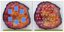
          We recently showed that in order to detect intra-tumor heterogeneity a Divide-and-Conquer (DAC) strategy of tumor sampling outperforms current routine protocols. This paper is a continuation of this work, but here we focus on DAC implementation in the Pathology Laboratory. In particular, we describe a new simple method that makes use of a cutting grid device and is applied to clear cell renal cell carcinomas for DAC implementation. This method assures a thorough sampling of large surgical specimens, facilitates the demonstration of intratumor heterogeneity, and saves time to pathologists in the daily practice. The method involves the following steps: 1. Thin slicing of the tumor (by hand or machine), 2. Application of a cutting grid to the slices ( e.g., a French fry cutter), resulting in multiple tissue cubes with fixed position within the slice, 3. Selection of tissue cubes for analysis, and finally, 4. Inclusion of selected cubes into a cassette for histological processing (with about eight tissue fragments within each cassette). Thus, using our approach in a 10 cm in-diameter-tumor we generate 80 tumor tissue fragments placed in 10 cassettes and, notably, in a tenth of time. Eighty samples obtained across all the regions of the tumor will assure a much higher performance in detecting intratumor heterogeneity, as proved recently with synthetic data.

          Related collections

          Most cited references9

          • Record: found
          • Abstract: found
          • Article: not found

          Handling and staging of renal cell carcinoma: the International Society of Urological Pathology Consensus (ISUP) conference recommendations.

          The International Society of Urologic Pathology 2012 Consensus Conference on renal cancer, through working group 3, focused on the issues of staging and specimen handling of renal tumors. The conference was preceded by an online survey of the International Society of Urologic Pathology members, and the results of this were used to inform the focus of conference discussion. On formal voting a ≥65% majority was considered a consensus agreement. For specimen handling it was agreed that with radical nephrectomy specimens the initial cut should be made along the long axis and that both radical and partial nephrectomy specimens should be inked. It was recommended that sampling of renal tumors should follow a general guideline of sampling 1 block/cm with a minimum of 3 blocks (subject to modification as needed in individual cases). When measuring a renal tumor, the length of a renal vein/caval thrombus should not be part of the measurement of the main tumor mass. In cases with multiple tumors, sampling should include at a minimum the 5 largest tumors. There was a consensus that perinephric fat invasion should be determined by examining multiple perpendicular sections of the tumor/perinephric fat interface and by sampling areas suspicious for invasion. Perinephric fat invasion was defined as either the tumor touching the fat or extending as irregular tongues into the perinephric tissue, with or without desmoplasia. It was agreed upon that renal sinus invasion is present when the tumor is in direct contact with the sinus fat or the loose connective tissue of the sinus, clearly beyond the renal parenchyma, or if there is involvement of any endothelium-lined spaces within the renal sinus, regardless of the size. When invasion of the renal sinus is uncertain, it was recommended that at least 3 blocks of the tumor-renal sinus interface should be submitted. If invasion is grossly evident, or obviously not present (small peripheral tumor), it was agreed that only 1 block was needed to confirm the gross impression. Other recommendations were that the renal vein margin be considered positive only when there is adherent tumor visible microscopically at the actual margin. When a specimen is submitted separately as "caval thrombus," the recommended sampling strategy is to take 2 or more sections to look for the adherent caval wall tissue. It was also recommended that uninvolved renal parenchyma be sampled by including normal parenchyma with tumor and normal parenchyma distant from the tumor. There was consensus that radical nephrectomy specimens should be examined for the purpose of identifying lymph nodes by dissection/palpation of the fat in the hilar area only; however, it was acknowledged that lymph nodes are found in <10% of radical nephrectomy specimens.
            Bookmark
            • Record: found
            • Abstract: not found
            • Article: not found

            Cell sorting: divide and conquer.

              Bookmark
              • Record: found
              • Abstract: found
              • Article: not found

              How should clinicians address intratumour heterogeneity in clear cell renal cell carcinoma?

              Despite the availability of multiple targeted therapies, the 5-year survival rate of patients with metastatic clear cell renal cell carcinoma (ccRCC) rarely exceeds 10%. Recent insights into the mutational landscape and evolutionary dynamics of ccRCC have offered up a plausible explanation for these outcomes. The purpose of this review is to link the research findings to potential changes in clinical practice.
                Bookmark

                Author and article information

                Journal
                F1000Res
                F1000Res
                F1000Research
                F1000Research
                F1000Research (London, UK )
                2046-1402
                26 July 2016
                2016
                : 5
                : 1587
                Affiliations
                [1 ]Department of Pathology, Cruces University Hospital, Barakaldo, Spain
                [2 ]Biomarkers in Cancer Unit, Biocruces Research Institute, Barakaldo, Spain
                [3 ]University of the Basque Country, Leioa, Spain
                [4 ]Quantitative Biomedicine Unit, Biocruces Research Institute, Barakaldo, Spain
                [5 ]Ikerbasque: The Basque Foundation for Science, Bilbao, Spain
                [6 ]Department of Cell Biology and Histology, University of the Basque Country, Leioa, Spain
                [1 ]Department of Laboratory Medicine and Pathology, Mayo Clinic, Scottsdale, AZ, USA
                [1 ]Department of Pathology, Brigham and Women's Hospital, Boston, MA, USA
                [1 ]Institute of Molecular Pathology and Immunology, University of Porto, Porto, Portugal
                [2 ]Department of Pathology and Oncology, University of Porto, Porto, Portugal
                [1 ]Department of Pathology, Brigham and Women's Hospital, Boston, MA, USA
                [1 ]Department of Laboratory Medicine and Pathology, Mayo Clinic, Scottsdale, AZ, USA
                Author notes

                JIL and JMC identified the problem and gave a realistic solution. JIL and JMC wrote this note.

                Competing interests: No competing interests were disclosed.

                Competing interests: No competing interests were disclosed.

                Competing interests: No competing interests were disclosed.

                Competing interests: No competing interests were disclosed.

                Competing interests: No competing interests were disclosed.

                Competing interests: No competing interests were disclosed.

                Article
                10.12688/f1000research.9091.2
                4965694
                27540472
                e52068af-de0f-494a-b5c4-7fd52bf6ee74
                Copyright: © 2016 Lopez JI and Cortes JM

                This is an open access article distributed under the terms of the Creative Commons Attribution Licence, which permits unrestricted use, distribution, and reproduction in any medium, provided the original work is properly cited.

                History
                : 25 July 2016
                Funding
                JMC is funded by Ikerbasque: The Basque Foundation for Science.
                The funders had no role in study design, data collection and analysis, decision to publish, or preparation of the manuscript.
                Categories
                Research Note
                Articles
                Genitourinary Cancers
                Methods for Diagnostic & Therapeutic Studies

                tumor sampling,cutting grid,divide and conquer,clear cell renal cell carcinoma,intratumor heterogeneity,pathology routine

                Comments

                Comment on this article