169
views
0
recommends
+1 Recommend
0 collections
    12
    shares
      • Record: found
      • Abstract: found
      • Article: found
      Is Open Access

      Morphometric, densitometric and mechanical properties of mandibular deciduous teeth in 5-month-old Polish Merino sheep

      research-article

      Read this article at

      Bookmark
          There is no author summary for this article yet. Authors can add summaries to their articles on ScienceOpen to make them more accessible to a non-specialist audience.

          Abstract

          Background

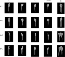
          Caries, enamel hypoplasia, molar incisor hipomineralization, amylogenesis imperfecta, dentine dysplasia, hypophosphatasia and other dental disorders lead to tooth mineralization disturbances and structural abnormalities, decreasing masticatory organ functions. Dental disorders in sheep can lead to premature slaughter before they have attained final stage of their reproductive life and induce economic loss due to high flock replacement costs. Growth rate, health status and meat quality of sheep depends on tooth properties and quality determining in large extent efficiency of the masticatory apparatus and initial food break up. Considering lack of basic anatomical and physiological data on teeth properties in sheep, the aim of the study was to evaluate morphometric, densitometric and mechanical traits of deciduous mandibular incisor, canine and the second premolar obtained at the slaughter age of 5 months of life.

          Results

          The obtained results have shown the highest values of weight, total tooth volume, enamel volume and dentine volume in second premolar. Morphometric and mechanical parameters of incisors reached the highest values in first incisor and decreased gradually in second and third incisor, and in canine. Densitometric measurements have not revealed significant differences of the volumetric tooth mineral density in hard dental tissues between the investigated teeth.

          Conclusions

          In conclusion, proposed methodological approach is noninvasive since the deciduous teeth undergo physiological replacement with permanent teeth. Deciduous teeth can be easy collected for analyses from large animal population and may reflect mineral status and metabolism resulting from postnatal growth and development of the whole flock. In individual cases, evaluation of properties of deciduous teeth may serve for breeding selection and further reproduction of sheep possessing favorable traits of teeth and better masticatory system functions.

          Related collections

          Most cited references21

          • Record: found
          • Abstract: found
          • Article: not found

          In vivo small-animal imaging using micro-CT and digital subtraction angiography.

          Small-animal imaging has a critical role in phenotyping, drug discovery and in providing a basic understanding of mechanisms of disease. Translating imaging methods from humans to small animals is not an easy task. The purpose of this work is to review in vivo x-ray based small-animal imaging, with a focus on in vivo micro-computed tomography (micro-CT) and digital subtraction angiography (DSA). We present the principles, technologies, image quality parameters and types of applications. We show that both methods can be used not only to provide morphological, but also functional information, such as cardiac function estimation or perfusion. Compared to other modalities, x-ray based imaging is usually regarded as being able to provide higher throughput at lower cost and adequate resolution. The limitations are usually associated with the relatively poor contrast mechanisms and potential radiation damage due to ionizing radiation, although the use of contrast agents and careful design of studies can address these limitations. We hope that the information will effectively address how x-ray based imaging can be exploited for successful in vivo preclinical imaging.
            Bookmark
            • Record: found
            • Abstract: found
            • Article: not found

            X-ray microtomographic study of mineral concentration distribution in deciduous enamel.

            To determine the mineral concentration distribution in deciduous enamel by quantitative X-ray microtomography (XMT). Tooth rods ( approximately 2 mm x 2 mm) were removed from the mid-buccal region of 11 deciduous molars. Three XMT slices were taken at 1.5, 2.0 and 2.5 mm from the amelocemental junction. The distribution and variation in mineral concentration of enamel were studied from the XMT images. The mean mineral concentration for all the teeth was 2.81 (S.D. = 0.065) g cm(-3). There was no notable difference in the mean mineral concentration values between the three XMT slices of each tooth. However, there was up to 8% variation between different teeth (2.69-2.92 g cm(-3)). Gradients of increasing mineral concentration from the amelodentinal junction (ADJ) to the external surface were found, ranging from 0.08 to 0.60 mg cm(-3) microm(-1) with a mean of 0.366 mg cm(-3) microm(-1). The mineral concentration gradients in the occlusal slices were steeper than those in the cervical slices. The difference in mineral concentration between the inner and outer enamel ranged from 1.5 to 8.7%. In view of the large variation in both the means and the gradients of mineral concentration in deciduous molars, the mineral distribution of each experimental tooth should be measured as baseline data in studies of caries progression.
              Bookmark
              • Record: found
              • Abstract: found
              • Article: not found

              Application of polychromatic µCT for mineral density determination.

              Accurate assessment of mineral density (MD) provides information critical to the understanding of mineralization processes of calcified tissues, including bones and teeth. High-resolution three-dimensional assessment of the MD of teeth has been demonstrated by relatively inaccessible synchrotron radiation microcomputed tomography (SRµCT). While conventional desktop µCT (CµCT) technology is widely available, polychromatic source and cone-shaped beam geometry confound MD assessment. Recently, considerable attention has been given to optimizing quantitative data from CµCT systems with polychromatic x-ray sources. In this review, we focus on the approaches that minimize inaccuracies arising from beam hardening, in particular, beam filtration during the scan, beam-hardening correction during reconstruction, and mineral density calibration. Filtration along with lowest possible source voltage results in a narrow and near-single-peak spectrum, favoring high contrast and minimal beam-hardening artifacts. More effective beam monochromatization approaches are described. We also examine the significance of beam-hardening correction in determining the accuracy of mineral density estimation. In addition, standards for the calibration of reconstructed grey-scale attenuation values against MD, including K(2)PHO(4) liquid phantom, and polymer-hydroxyapatite (HA) and solid hydroxyapatite (HA) phantoms, are discussed.
                Bookmark

                Author and article information

                Journal
                BMC Vet Res
                BMC Vet. Res
                BMC Veterinary Research
                BioMed Central
                1746-6148
                2014
                19 February 2014
                : 10
                : 45
                Affiliations
                [1 ]Department of Animal Physiology, Faculty of Veterinary Medicine, University of Life Sciences in Lublin, ul. Akademicka 12, 20-950 Lublin Poland
                [2 ]II Department of Radiology, Medical University in Lublin, ul. Staszica 16, 20-081 Lublin, Poland
                [3 ]Department of Prosthetic Dentistry, Medical University in Lublin, ul. Karmelicka 7, 20-081 Lublin, Poland
                [4 ]Department of Conservative Dentistry and Endodontics, Medical University in Lublin, ul. Karmelicka 7, 20-081 Lublin, Poland
                [5 ]Department of Animal Anatomy, Faculty of Veterinary Medicine, University of Life Sciences in Lublin, ul. Akademicka 12, 20-950 Lublin, Poland
                [6 ]Department of Materials Engineering, Lublin University of Technology, ul. Nadbystrzycka 36, 20-618 Lublin, Poland
                Article
                1746-6148-10-45
                10.1186/1746-6148-10-45
                3936944
                24548814
                e57db2cc-dfb6-4b37-a868-a9551f50a821
                Copyright © 2014 Tatara et al.; licensee BioMed Central Ltd.

                This is an Open Access article distributed under the terms of the Creative Commons Attribution License ( http://creativecommons.org/licenses/by/2.0), which permits unrestricted use, distribution, and reproduction in any medium, provided the original work is properly credited.

                History
                : 16 March 2013
                : 15 February 2014
                Categories
                Research Article

                Veterinary medicine
                densitometry,teeth,quantitative computed tomography,microtomography,sheep
                Veterinary medicine
                densitometry, teeth, quantitative computed tomography, microtomography, sheep

                Comments

                Comment on this article