1
views
0
recommends
+1 Recommend
0 collections
    0
    shares
      • Record: found
      • Abstract: found
      • Article: found
      Is Open Access

      Type 2 Diabetic Mellitus Inhibits Skin Renewal through Inhibiting WNT-Dependent Lgr5+ Hair Follicle Stem Cell Activation in C57BL/6 Mice

      research-article

      Read this article at

      Bookmark
          There is no author summary for this article yet. Authors can add summaries to their articles on ScienceOpen to make them more accessible to a non-specialist audience.

          Abstract

          Background

          Hair follicles are important accessory organs of the skin, and it is important for skin renewal and performs variety of important functions. Diabetes can cause several dermatoses; however, its effect on hair follicles is unclear. The purpose of this study was to investigate the effect of type II diabetes (T2DM) on the hair follicles of mice.

          Methods

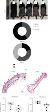
          Seven-week-old male C57BL/6 littermate mice were divided into two groups. The treatment group was injected with streptozotocin (STZ) to induce T2DM, and the control group was parallelly injected with the same dose of buffer. Seven days after injection, the back is depilated to observe the hair follicle regeneration. Hair follicle regeneration was observed by naked eyes and HE staining. The proliferation of the skin cells was observed by PCNA and K14 staining. The altered genes were screened by RNA sequencing and verified by qRT-PCR. In addition, Lgr5 + GFP/mTmG transgenic mice were used to observe the effect of T2DM on Lgr5 hair follicle stem cells (HFSC). And the expression of WNT4 and WNT8A were measured by Western Blot.

          Results

          T2DM inhibited hair follicle regeneration. Compared to control mice, T2DM mice had smaller hair follicles, reduced skin thickness, and less expression of PCNA and K14. RNA sequencing showed that the two groups had significant differences in cell cycle and proliferation-related pathways. Compared with the control mice, the mRNA expression of Lgr4, Lgr5, Wnt4, and Wnt8a was decreased in the T2DM group. Moreover, T2DM inhibited the activation of Lgr5 HFSC and the expression of WNT4 and WNT8A.

          Conclusions

          T2DM inhibited hair follicle regeneration and skin cells proliferation by inhibiting WNT-dependent Lgr5 HFSC activation. This may be an important reason for the reduction of skin renewal ability and the formation of chronic wounds caused by diabetes. It is important for the treatment of chronic diabetic wounds and the development of tissue engineering.

          Related collections

          Most cited references54

          • Record: found
          • Abstract: found
          • Article: not found

          Identification of stem cells in small intestine and colon by marker gene Lgr5.

          The intestinal epithelium is the most rapidly self-renewing tissue in adult mammals. It is currently believed that four to six crypt stem cells reside at the +4 position immediately above the Paneth cells in the small intestine; colon stem cells remain undefined. Lgr5 (leucine-rich-repeat-containing G-protein-coupled receptor 5, also known as Gpr49) was selected from a panel of intestinal Wnt target genes for its restricted crypt expression. Here, using two knock-in alleles, we reveal exclusive expression of Lgr5 in cycling columnar cells at the crypt base. In addition, Lgr5 was expressed in rare cells in several other tissues. Using an inducible Cre knock-in allele and the Rosa26-lacZ reporter strain, lineage-tracing experiments were performed in adult mice. The Lgr5-positive crypt base columnar cell generated all epithelial lineages over a 60-day period, suggesting that it represents the stem cell of the small intestine and colon. The expression pattern of Lgr5 suggests that it marks stem cells in multiple adult tissues and cancers.
            Bookmark
            • Record: found
            • Abstract: found
            • Article: not found

            Global epidemiology of diabetic foot ulceration: a systematic review and meta-analysis †.

            Diabetic foot is a severe public health issue, yet rare studies investigated its global epidemiology. Here we performed a systematic review and meta-analysis through searching PubMed, EMBASE, ISI Web of science, and Cochrane database. We found that that global diabetic foot ulcer prevalence was 6.3% (95%CI: 5.4-7.3%), which was higher in males (4.5%, 95%CI: 3.7-5.2%) than in females (3.5%, 95%CI: 2.8-4.2%), and higher in type 2 diabetic patients (6.4%, 95%CI: 4.6-8.1%) than in type 1 diabetics (5.5%, 95%CI: 3.2-7.7%). North America had the highest prevalence (13.0%, 95%CI: 10.0-15.9%), Oceania had the lowest (3.0%, 95% CI: 0.9-5.0%), and the prevalence in Asia, Europe, and Africa were 5.5% (95%CI: 4.6-6.4%), 5.1% (95%CI: 4.1-6.0%), and 7.2% (95%CI: 5.1-9.3%), respectively. Australia has the lowest (1.5%, 95%CI: 0.7-2.4%) and Belgium has the highest prevalence (16.6%, 95%CI: 10.7-22.4%), followed by Canada (14.8%, 95%CI: 9.4-20.1%) and USA (13.0%, 95%CI: 8.3-17.7%). The patients with diabetic foot ulcer were older, had a lower body mass index, longer diabetic duration, and had more hypertension, diabetic retinopathy, and smoking history than patients without diabetic foot ulceration. Our results provide suggestions for policy makers in deciding preventing strategy of diabetic foot ulceration in the future. Key messages Global prevalence of diabetic foot is 6.3% (95%CI: 5.4-7.3%), and the prevalence in North America, Asia, Europe, Africa and Oceania was 13.0% (95%CI: 10.0-15.9%), 5.5% (95%CI: 4.6-6.4%), 5.1% (95%CI: 4.1-6.0%), 7.2% (95%CI: 5.1-9.3%), and 3.0% (95% CI: 0.9-5.0%). Diabetic foot was more prevalent in males than in females, and more prevalent in type 2 diabetic foot patients than in type 1 diabetic foot patients. The patients with diabetic foot were older, had a lower body mass index, longer diabetic duration, and had more hypertension, diabetic retinopathy, and smoking history than patients without diabetic foot.
              Bookmark
              • Record: found
              • Abstract: not found
              • Article: not found
              Is Open Access

              Mechanistic insight into diabetic wounds: Pathogenesis, molecular targets and treatment strategies to pace wound healing

                Bookmark

                Author and article information

                Contributors
                Journal
                J Diabetes Res
                J Diabetes Res
                JDR
                Journal of Diabetes Research
                Hindawi
                2314-6745
                2314-6753
                2022
                16 April 2022
                : 2022
                : 8938276
                Affiliations
                1Department of Burn and Plastic Surgery, Shenzhen Institute of Translational Medicine, Shenzhen Second People's Hospital, The First Affiliated Hospital of Shenzhen University Health Science Center, Shenzhen 518035, China
                2Human Histology & Embryology Section, Department of Surgery, Dentistry, Pediatrics & Gynecology, University of Verona Medical School, Verona 37134, Italy
                Author notes

                Academic Editor: Ali Sharif

                Author information
                https://orcid.org/0000-0002-4993-6606
                https://orcid.org/0000-0002-4997-1970
                https://orcid.org/0000-0001-5271-7079
                Article
                10.1155/2022/8938276
                9034921
                35469170
                e5d67bd5-7d4f-4066-ab8d-68022673e889
                Copyright © 2022 Minghui Wang et al.

                This is an open access article distributed under the Creative Commons Attribution License, which permits unrestricted use, distribution, and reproduction in any medium, provided the original work is properly cited.

                History
                : 23 November 2021
                : 24 March 2022
                : 25 March 2022
                Funding
                Funded by: Shenzhen Sanming Project
                Award ID: 30000013
                Funded by: Shenzhen Postdoctoral Research Grant
                Award ID: 50820191286
                Funded by: China Postdoctoral Science Foundation
                Award ID: 2018M630141
                Categories
                Research Article

                Comments

                Comment on this article