2
views
0
recommends
+1 Recommend
1 collections
    0
    shares
      • Record: found
      • Abstract: found
      • Article: found
      Is Open Access

      Shikonin promotes hypertrophic scar repair by autophagy of hypertrophic scar-derived fibroblasts

      research-article

      Read this article at

      ScienceOpenPublisherPMC
      Bookmark
          There is no author summary for this article yet. Authors can add summaries to their articles on ScienceOpen to make them more accessible to a non-specialist audience.

          ABSTRACT

          Purpose:

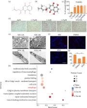
          To investigate the Shikonin (SHI) induce autophagy of hypertrophic scar-derived fibroblasts (HSFs) and the mechanism of which in repairing hypertrophic scar.

          Methods:

          This study showed that SHI induced autophagy from HSFs and repaired skin scars through the AMPK/mTOR pathway. Alamar Blue and Sirius red were used to identify cell activity and collagen. Electron microscopy, label-free quantitative proteomic analysis, fluorescence and other methods were used to identify autophagy. The differences in the expression of autophagy and AMPK/mTOR pathway-related proteins after SHI treatment were quantitatively analyzed by Western blots. A quantitative real-time polymerase chain reaction assay was used to detect the expression of LC3, AMPK and ULK after adding chloroquine (CQ) autophagy inhibitor.

          Results:

          After treatment with SHI for 24 hours, it was found that the viability of HSFs was significantly reduced, the protein expression of LC3-II/LC3-I and Beclin1 increased, while the protein expression of P62 decreased. The expression of phosphorylated AMPK increased and expression of phosphorylated mTOR decreased. After the use of CQ, the cell autophagy caused by SHI was blocked. The key genes LC3 and P62 were then reexamined by immunohistochemistry using a porcine full-thickness burn hypertrophic scar model, and the results verified that SHI could induce autophagy in vivo.

          Conclusions:

          These findings suggested that SHI promoted autophagy of HSFs cells, and the potential mechanism may be related to the AMPK/mTOR signal pathway, which provided new insights for the treatment of hypertrophic scars.

          Related collections

          Most cited references44

          • Record: found
          • Abstract: found
          • Article: not found

          AMPK and mTOR regulate autophagy through direct phosphorylation of Ulk1.

          Autophagy is a process by which components of the cell are degraded to maintain essential activity and viability in response to nutrient limitation. Extensive genetic studies have shown that the yeast ATG1 kinase has an essential role in autophagy induction. Furthermore, autophagy is promoted by AMP activated protein kinase (AMPK), which is a key energy sensor and regulates cellular metabolism to maintain energy homeostasis. Conversely, autophagy is inhibited by the mammalian target of rapamycin (mTOR), a central cell-growth regulator that integrates growth factor and nutrient signals. Here we demonstrate a molecular mechanism for regulation of the mammalian autophagy-initiating kinase Ulk1, a homologue of yeast ATG1. Under glucose starvation, AMPK promotes autophagy by directly activating Ulk1 through phosphorylation of Ser 317 and Ser 777. Under nutrient sufficiency, high mTOR activity prevents Ulk1 activation by phosphorylating Ulk1 Ser 757 and disrupting the interaction between Ulk1 and AMPK. This coordinated phosphorylation is important for Ulk1 in autophagy induction. Our study has revealed a signalling mechanism for Ulk1 regulation and autophagy induction in response to nutrient signalling.
            Bookmark
            • Record: found
            • Abstract: found
            • Article: not found

            Autophagy: renovation of cells and tissues.

            Autophagy is the major intracellular degradation system by which cytoplasmic materials are delivered to and degraded in the lysosome. However, the purpose of autophagy is not the simple elimination of materials, but instead, autophagy serves as a dynamic recycling system that produces new building blocks and energy for cellular renovation and homeostasis. Here we provide a multidisciplinary review of our current understanding of autophagy's role in metabolic adaptation, intracellular quality control, and renovation during development and differentiation. We also explore how recent mouse models in combination with advances in human genetics are providing key insights into how the impairment or activation of autophagy contributes to pathogenesis of diverse diseases, from neurodegenerative diseases such as Parkinson disease to inflammatory disorders such as Crohn disease. Copyright © 2011 Elsevier Inc. All rights reserved.
              Bookmark
              • Record: found
              • Abstract: found
              • Article: not found

              p62/SQSTM1 binds directly to Atg8/LC3 to facilitate degradation of ubiquitinated protein aggregates by autophagy.

              Protein degradation by basal constitutive autophagy is important to avoid accumulation of polyubiquitinated protein aggregates and development of neurodegenerative diseases. The polyubiquitin-binding protein p62/SQSTM1 is degraded by autophagy. It is found in cellular inclusion bodies together with polyubiquitinated proteins and in cytosolic protein aggregates that accumulate in various chronic, toxic, and degenerative diseases. Here we show for the first time a direct interaction between p62 and the autophagic effector proteins LC3A and -B and the related gamma-aminobutyrate receptor-associated protein and gamma-aminobutyrate receptor-associated-like proteins. The binding is mediated by a 22-residue sequence of p62 containing an evolutionarily conserved motif. To monitor the autophagic sequestration of p62- and LC3-positive bodies, we developed a novel pH-sensitive fluorescent tag consisting of a tandem fusion of the red, acid-insensitive mCherry and the acid-sensitive green fluorescent proteins. This approach revealed that p62- and LC3-positive bodies are degraded in autolysosomes. Strikingly, even rather large p62-positive inclusion bodies (2 microm diameter) become degraded by autophagy. The specific interaction between p62 and LC3, requiring the motif we have mapped, is instrumental in mediating autophagic degradation of the p62-positive bodies. We also demonstrate that the previously reported aggresome-like induced structures containing ubiquitinated proteins in cytosolic bodies are dependent on p62 for their formation. In fact, p62 bodies and these structures are indistinguishable. Taken together, our results clearly suggest that p62 is required both for the formation and the degradation of polyubiquitin-containing bodies by autophagy.
                Bookmark

                Author and article information

                Contributors
                Role: Substantive scientific and intellectual contributions to the studyRole: Conception and designRole: Analysis and interpretation of dataRole: Statistics analysisRole: Manuscript preparationRole: Manuscript writing
                Role: Acquisition of dataRole: Analysis and interpretation of dataRole: Manuscript preparation
                Role: Acquisition of dataRole: Histopathological examinationsRole: Critical revision
                Role: Analysis and interpretation of dataRole: Statistics analysis
                Role: Analysis and interpretation of dataRole: Statistics analysis
                Role: Analysis and interpretation of data
                Role: Analysis and interpretation of data
                Role: Analysis and interpretation of data
                Journal
                Acta Cir Bras
                Acta Cir Bras
                acta
                Acta Cirúrgica Brasileira
                Sociedade Brasileira para o Desenvolvimento da Pesquisa em Cirurgia
                0102-8650
                1678-2674
                23 October 2023
                2023
                : 38
                : e384623
                Affiliations
                [1 ]Ningxia Medical University General Hospital – Tissue Organ Bank & Tissue Engineering Centre – c (Ningxia) – China.
                [2 ]Ningxia Medical University – Key Laboratory of Fertility Preservation and Maintenance of Ministry of Education – School of Basic Medicine – Yinchuan (Ningxia) – China.
                [3 ]Ningxia Medical University – Clinical Medical School – Yinchuan (Ningxia) – China
                [4 ]The First People’s Hospital – Department of Burns and Plastic Surgery – Shizuishan – China.
                Author notes
                [* ]Corresponding author: 516790015@ 123456qq.com

                About the authors: Zhang Q and Zhao D are PhD students.

                Wang M, Deng X, Zhao F and Xiao J are Master’s degree students.

                Ma J and Pan X are Undergraduate Students.

                Conflict of interest: Nothing to declare.

                Author information
                http://orcid.org/0000-0002-9136-7718
                http://orcid.org/0000-0002-5066-728X
                http://orcid.org/0000-0001-8080-7231
                http://orcid.org/0000-0003-3834-0142
                http://orcid.org/0000-0002-9655-0538
                http://orcid.org/0000-0002-2601-2400
                http://orcid.org/0000-0002-1121-5192
                http://orcid.org/0009-0002-8172-0212
                Article
                00237
                10.1590/acb384623
                10592587
                e6dbb29d-53dc-4896-95e8-d02db9c9e8de

                This is an Open Access article distributed under the terms of the Creative Commons Attribution License, which permits unrestricted use, distribution, and reproduction in any medium, provided the original work is properly cited.

                History
                : 23 May 2023
                : 14 August 2023
                Page count
                Figures: 3, Tables: 2, Equations: 1, References: 37
                Categories
                Original Article

                shikonin,autophagy,cicatrix
                shikonin, autophagy, cicatrix

                Comments

                Comment on this article