94
views
0
recommends
+1 Recommend
0 collections
    0
    shares
      • Record: found
      • Abstract: found
      • Article: not found

      Functional Organization of Human Sensorimotor Cortex for Speech Articulation

      research-article

      Read this article at

      ScienceOpenPublisherPMC
      Bookmark
          There is no author summary for this article yet. Authors can add summaries to their articles on ScienceOpen to make them more accessible to a non-specialist audience.

          Abstract

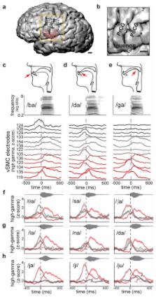
          Speaking is one of the most complex actions we perform, yet nearly all of us learn to do it effortlessly. Production of fluent speech requires the precise, coordinated movement of multiple articulators (e.g., lips, jaw, tongue, larynx) over rapid time scales. Here, we used high-resolution, multi-electrode cortical recordings during the production of consonant-vowel syllables to determine the organization of speech sensorimotor cortex in humans. We found speech articulator representations that were somatotopically arranged on ventral pre- and post-central gyri and partially overlapping at individual electrodes. These representations were temporally coordinated as sequences during syllable production. Spatial patterns of cortical activity revealed an emergent, population-level representation, which was organized by phonetic features. Over tens of milliseconds, the spatial patterns transitioned between distinct representations for different consonants and vowels. These results reveal the dynamic organization of speech sensorimotor cortex during the generation of multi-articulator movements underlying our ability to speak.

          Related collections

          Most cited references47

          • Record: found
          • Abstract: found
          • Article: not found

          Neural population dynamics during reaching

          Most theories of motor cortex have assumed that neural activity represents movement parameters. This view derives from an analogous approach to primary visual cortex, where neural activity represents patterns of light. Yet it is unclear how well that analogy holds. Single-neuron responses in motor cortex appear strikingly complex, and there is marked disagreement regarding which movement parameters are represented. A better analogy might be with other motor systems, where a common principle is rhythmic neural activity. We found that motor cortex responses during reaching contain a brief but strong oscillatory component, something quite unexpected for a non-periodic behavior. Oscillation amplitude and phase followed naturally from the preparatory state, suggesting a mechanistic role for preparatory neural activity. These results demonstrate unexpected yet surprisingly simple structure in the population response. That underlying structure explains many of the confusing features of individual-neuron responses.
            Bookmark
            • Record: found
            • Abstract: found
            • Article: not found

            Generating coherent patterns of activity from chaotic neural networks.

            Neural circuits display complex activity patterns both spontaneously and when responding to a stimulus or generating a motor output. How are these two forms of activity related? We develop a procedure called FORCE learning for modifying synaptic strengths either external to or within a model neural network to change chaotic spontaneous activity into a wide variety of desired activity patterns. FORCE learning works even though the networks we train are spontaneously chaotic and we leave feedback loops intact and unclamped during learning. Using this approach, we construct networks that produce a wide variety of complex output patterns, input-output transformations that require memory, multiple outputs that can be switched by control inputs, and motor patterns matching human motion capture data. Our results reproduce data on premovement activity in motor and premotor cortex, and suggest that synaptic plasticity may be a more rapid and powerful modulator of network activity than generally appreciated.
              Bookmark
              • Record: found
              • Abstract: found
              • Article: not found

              Functional mapping of human sensorimotor cortex with electrocorticographic spectral analysis. II. Event-related synchronization in the gamma band.

              N Crone (1998)
              It has been shown in animals that neuronal activity in the 'gamma band' (>30 Hz) is associated with cortical activation and may play a role in multi-regional and multi-modal integration of cortical processing. Studies of gamma activity in human scalp EEG have typically focused on event-related synchronization (ERS) in the 40 Hz band. To assess further the gamma band ERS further, as an index of cortical activation and as a tool for human functional brain mapping, we recorded subdural electrocorticographic (ECoG) signals in five clinical subjects while they performed visual-motor decision tasks designed to activate the representations of different body parts in sensorimotor cortex. ECoG spectral analysis utilized a mixed-effects analysis of variance model in which within-trial temporal dependencies were accounted for. Taking an exploratory approach, we studied gamma ERS in 10-Hz-wide bands (overlapping by 5 Hz) ranging from 30 to 100 Hz, and compared these findings with changes in the alpha (8-13 Hz) and beta (15-25 Hz) bands. Gamma ERS (observed in three out of subjects) occurred in two broad bands-'low gamma' included the 35-45 and 40-50 Hz bands, and 'high gamma' the 75-85, 80-90, 85-95 and 90-100 Hz bands. The temporal and spatial characteristics of low and high gamma ERS were distinct, suggesting relatively independent neurophysiological mechanisms. Low gamma ERS often began after onset of the motor response and was sustained through much of it, in parallel with event-related desynchronization (ERD) in the alpha band. High gamma ERS often began during, or slightly before, the motor response and was transient, ending well before completion of the motor response. These temporal differences in low and high gamma suggest different functional associations with motor performance. Compared with alpha and beta ERD, the topographical patterns of low and high gamma ERS were more discrete and somatotopically specific and only occurred over contralateral sensorimotor cortex during unilateral limb movements (alpha and beta ERD were also observed ipsilaterally). Maps of sensorimotor function inferred from gamma ERS were consistent with maps generated by cortical electrical stimulation for clinical purposes. In addition, different task conditions in one subject produced consistent differences in both motor response latencies and onset latency of gamma ERS, particularly high gamma ERS. Compared with alpha and beta ERD, the topography of gamma ERS is more consistent with traditional maps of sensorimotor functional anatomy. In addition, gamma ERS may provide complementary information about cortical neurophysiology that is useful for mapping brain function in humans.
                Bookmark

                Author and article information

                Journal
                0410462
                6011
                Nature
                Nature
                Nature
                0028-0836
                1476-4687
                24 January 2013
                20 February 2013
                21 March 2013
                21 September 2013
                : 495
                : 7441
                : 327-332
                Affiliations
                [1 ]Departments of Neurological Surgery and Physiology, University of California, San Francisco
                [2 ]Center for Integrative Neuroscience, University of California, San Francisco
                [3 ]Department of Linguistics, University of California, Berkeley
                [4 ]UCSF Epilepsy Center, University of California, San Francisco
                Author notes
                Correspondence and requests for materials should be addressed to E.F.C. ( ChangEd@ 123456neurosurg.ucsf.edu )
                Article
                NIHMS436351
                10.1038/nature11911
                3606666
                23426266
                e6ea1610-48d4-4947-b56d-4803797eeda0

                Users may view, print, copy, download and text and data- mine the content in such documents, for the purposes of academic research, subject always to the full Conditions of use: http://www.nature.com/authors/editorial_policies/license.html#terms

                History
                Funding
                Funded by: National Institute on Deafness and Other Communication Disorders : NIDCD
                Award ID: R01 DC012379 || DC
                Funded by: National Institute of Neurological Disorders and Stroke : NINDS
                Award ID: R00 NS065120 || NS
                Funded by: National Institute of Neurological Disorders and Stroke : NINDS
                Award ID: L30 NS060463 || NS
                Funded by: Office of the Director : NIH
                Award ID: DP2 OD008627 || OD
                Categories
                Article

                Uncategorized
                Uncategorized

                Comments

                Comment on this article