3
views
0
recommends
+1 Recommend
0 collections
    0
    shares
      • Record: found
      • Abstract: found
      • Article: found
      Is Open Access

      Retinal pigment epithelium aperture: A late-onset complication in adult-onset foveomacular vitelliform dystrophy

      research-article

      Read this article at

      Bookmark
          There is no author summary for this article yet. Authors can add summaries to their articles on ScienceOpen to make them more accessible to a non-specialist audience.

          Abstract

          Purpose:

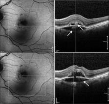
          The purpose of the study was to report aperture of retinal pigment epithelium (RPE) as a late complication and an unreported finding during the natural course of adult-onset foveomacular vitelliform dystrophy (AFVD).

          Methods:

          Four diagnosed cases of AFVD followed for a period ranging from 4 to 8 years. All patients had documented records of clinical examination, fundus autofluorescence and fluorescein angiography, and spectral domain-optical coherence tomography at regular intervals.

          Results:

          Besides the known stages in the natural course of AFVD, RPE aperture was noted as an additional finding during the vitelliruptive stage of the disease. The vitelliform material was noted beneath the disrupted RPE before disappearance. Accumulation of vitelliform material continued even after the vitelliruptive stage.

          Conclusion:

          RPE aperture may represent an ongoing process in the natural course of AFVD.

          Related collections

          Most cited references18

          • Record: found
          • Abstract: found
          • Article: not found

          Histologic and Optical Coherence Tomographic Correlates in Drusenoid Pigment Epithelium Detachment in Age-Related Macular Degeneration.

          Drusenoid pigment epithelium detachment (DPED) is a known precursor to geographic atrophy in age-related macular degeneration (AMD). We sought histologic correlates for spectral-domain (SD) optical coherence tomography (OCT) signatures in DPED and determined the frequency and origin of these OCT signatures in a clinical cohort of DPED eyes.
            Bookmark
            • Record: found
            • Abstract: found
            • Article: not found

            Intraretinal Hyperreflective Foci in Acquired Vitelliform Lesions of the Macula: Clinical and Histologic Study.

            To describe the natural course, visual outcomes, and anatomic changes and provide histologic correlates in eyes with intraretinal hyperreflective foci associated with acquired vitelliform lesions.
              Bookmark
              • Record: found
              • Abstract: found
              • Article: not found

              Acquired Vitelliform Lesions: correlation of clinical findings and multiple imaging analyses.

              To correlate clinical observations with multimodal imaging analysis in acquired vitelliform lesions (AVLs) and to elucidate their nature, pathogenesis, and natural course. Clinical examination, color fundus photography, fluorescein angiography, near-infrared reflectance, fundus autofluorescence, and spectral-domain optical coherence tomography (SD-OCT) data were retrospectively reviewed for a consecutive series of 90 eyes of 67 patients with an AVL secondary to a variety of diagnoses. In all 90 eyes with AVLs, SD-OCT helped localize the clinically apparent yellowish material to the subretinal space above the retinal pigment epithelial (RPE) band. Only 19 eyes (21.1%) had SD-OCT evidence of subretinal fluid. All eyes exhibited abnormal hyperautofluorescence corresponding to the material seen clinically. In 18 of 72 eyes (25.0%) imaged simultaneously with SD-OCT and near-infrared reflectance imaging, near-infrared hyperreflectivity corresponding to presumed collections of pigment-laden macrophages and RPE cells was observed. Resolution of some AVLs appeared to coincide with progressive thinning of the outer nuclear layer, indicating a gradual loss of photoreceptors. Visual acuity at baseline was best predicted by the subfoveal integrity of the external limiting membrane (P = 0.001) and the inner segment/outer segment junction (P = 0.0002) on SD-OCT. Fluorescein angiography revealed a pattern often mimicking poorly defined (Type 1) choroidal neovascularization. Acquired vitelliform lesions occur in a variety of different clinical entities that share common features with multimodal imaging analyses. We propose that both dysfunctional RPE and loss of apposition between the photoreceptor tips and the RPE can interfere with the phagocytosis of shed outer segments. Both this material and pigment-laden macrophages and RPE cells appear to contribute to the yellowish appearance of AVLs. Ongoing photoreceptor loss may in some cases be associated with the spontaneous resolution of an AVL.
                Bookmark

                Author and article information

                Journal
                Indian J Ophthalmol
                Indian J Ophthalmol
                IJO
                Indian Journal of Ophthalmology
                Medknow Publications & Media Pvt Ltd (India )
                0301-4738
                1998-3689
                January 2018
                : 66
                : 1
                : 83-88
                Affiliations
                [1]Advanced Eye Centre, Post Graduate Institute of Medical Education and Research, Chandigarh, India
                Author notes
                Correspondence to: Dr. Amod Gupta, Advanced Eye Centre, Post Graduate Institute of Medical Education and Research, Chandigarh, India. E-mail: dramodgupta@ 123456gmail.com
                Article
                IJO-66-83
                10.4103/ijo.IJO_676_17
                5778589
                29283129
                e95dfb4a-72de-4e7b-a0d9-98ec120234ab
                Copyright: © 2017 Indian Journal of Ophthalmology

                This is an open access article distributed under the terms of the Creative Commons Attribution-NonCommercial-ShareAlike 3.0 License, which allows others to remix, tweak, and build upon the work non-commercially, as long as the author is credited and the new creations are licensed under the identical terms.

                History
                : 01 August 2017
                : 03 October 2017
                Categories
                Original Article

                Ophthalmology & Optometry
                adult-onset foveomacular vitelliform dystrophy,aperture,fundus autofluorescence,retinal pigment epithelium,spectral domain-optical coherence tomography

                Comments

                Comment on this article