10
views
0
recommends
+1 Recommend
0 collections
    0
    shares
      • Record: found
      • Abstract: found
      • Article: not found

      Crystalline lens changes in porcine eyes with implanted phakic IOL (ICL) with a central hole

      research-article

      Read this article at

      ScienceOpenPublisherPMC
      Bookmark
          There is no author summary for this article yet. Authors can add summaries to their articles on ScienceOpen to make them more accessible to a non-specialist audience.

          Abstract

          Background

          We calculated the smallest diameter of a hole in the center of the optic at which the optical character of a phakic IOL (ICL) may be maintained. The changes induced in the aqueous humor dynamics and the pathology of cataract development with such a hole were investigated.

          Methods

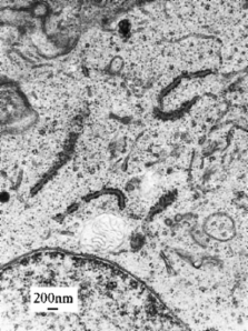
          A simulation was performed using ZEMAX software to calculate the hole diameter that makes possible the maintenance of a stable optical character of a phakic IOL. After a hole of calculated diameter was trepanned in the center of the optic of the ICL, the latter was implanted into one eye of a 5-month-old minipig, and an unperforated ICL into the other. The postoperative course was observed for 3 months. Then, Evans blue was injected into the vitreous body under general anesthesia to stain the anterior capsule of the crystalline lens. Within 30 min, the eye was enucleated and the tissues removed were fixed.

          Results

          The MTF of the perforated ICL (hole diameter, 1.0 mm) in the center of the optic resembled that of the unperforated ICL. In all cases with non-perforated ICLs, subcapsular turbidity developed, but no staining caused by EB was observed in the anterior capsule. On the other hand, the anterior capsules of the eyes fitted with ICLs with a 1.0-mm hole were stained, but exhibited no turbidity.

          Conclusion

          An ICL with a central hole of diameter 1.0 mm in the optic is similar to an unperforated ICL. The size of the hole influenced the aqueous humor dynamics and increased the aqueous humor perfusion volume over the entire anterior surface of the crystalline lens. The possibility of preventing cataracts was therefore suggested.

          Related collections

          Most cited references19

          • Record: found
          • Abstract: found
          • Article: not found

          United States Food and Drug Administration clinical trial of the Implantable Collamer Lens (ICL) for moderate to high myopia: three-year follow-up.

          To report on 3-year postoperative safety and efficacy outcomes with the Myopic Implantable Collamer Lens (ICL). Prospective nonrandomized clinical trial. Five hundred twenty-six eyes of 294 patients with between 3.0 and 20.0 diopters (D) of myopia participating in the United States Food and Drug Administration clinical trial of the ICL for myopia. Implantation of the ICL. Uncorrected visual acuity (VA), refraction, best spectacle-corrected VA (BSCVA), adverse events, operative and postoperative complications, lens opacity analysis, subjective satisfaction, and patient symptoms. At 3 years, 59.3% had 20/20 or better VA, and 94.7% had 20/40 or better uncorrected VA if BSCVA was 20/20 and patients were targeted for emmetropia; 67.5% of patients were within 0.5 D and 88.2% were within 1.0 D of predicted refraction. The mean improvement in BSCVA ranged between 0.5 and 0.6 lines. At 3 years postoperatively, 3 eyes (0.8%) decreased by >or=2 lines of BSCVA, in contrast to 40 eyes (10.8%) that improved by a similar amount. Contrast sensitivity improved postoperatively. Cumulative 3-year corneal endothelial cell loss was under 10%. Early largely asymptomatic, presumably surgically induced anterior subcapsular opacities (trace or greater) were seen in 14 eyes (2.7%), with only 2 being clinically significant. Five eyes (0.9%) of 3 patients developed nuclear opacities of grade >2 at 2 to 3 years postoperatively. Three (0.6%) ICL removals with cataract extraction and IOL implantation have been performed. Only 0.6% reported dissatisfaction; 97.1% of patients reported they would choose ICL implantation again. Incidences of patient symptoms, glare, halos, double vision, night vision problems, and night driving difficulties decreased or remained unchanged after ICL surgery. Three-year results from this standardized, multicenter clinical investigation support the safety, efficacy, and predictability of ICL surgery to treat moderate to high myopic refractive errors.
            Bookmark
            • Record: found
            • Abstract: found
            • Article: not found

            U.S. Food and Drug Administration clinical trial of the Implantable Contact Lens for moderate to high myopia.

            (2003)
            To assess the safety and efficacy of the Implantable Contact Lens (ICL) to treat moderate to high myopia. Prospective nonrandomized clinical trial. Five hundred twenty-three eyes of 291 patients with between 3 and 20.0 diopters (D) of myopia participating in the U. S. Food and Drug Administration clinical trial of the ICL for myopia. Implantation of the ICL. Uncorrected visual acuity (UCVA), refraction, best spectacle-corrected visual acuity (BSCVA), adverse events, operative and postoperative complications, lens opacity analysis (Lens Opacity Classification System III), subjective satisfaction, and symptoms. Twelve months postoperatively, 60.1% of patients had a visual acuity of 20/20 or better, and 92.5% had an uncorrected visual acuity of 20/40 or better. Patients averaged a 10.31-line improvement in UCVA, 61.6% of patients were within 0.5 D, and 84.7% were within 1.0 D of predicted refraction. Only one case (0.2%) lost > 2 lines of BSCVA. Gains of 2 or more lines of BSCVA occurred in 55 cases (11.8%) at 6 months and 41 cases (9.6%) at 1 year after ICL surgery. Early and largely asymptomatic, presumably surgically induced anterior subcapsular (AS) opacities were seen in 11 cases (2.1%); an additional early AS opacity (0.2%) was seen because of inadvertent anterior chamber irrigation of preservative-containing solution at surgery. Two (0.4%) late (> or = 1 year postoperatively) AS opacities were observed. Two (0.4%) ICL removals with cataract extraction and intraocular lens implantation have been performed. Patient satisfaction (very/extremely satisfied) was reported by 92.4% of subjects on the subjective questionnaire; only four patients (1.0%) reported dissatisfaction. Slightly more patients reported an improvement at 1 year over baseline values for the following subjective symptoms: quality of vision, glare, double vision, and night driving difficulties. Only a 3% difference between pre-ICL and post-ICL surgery was reported for haloes. The results support the safety, efficacy, and predictability of ICL implantation to treat moderate to high myopia.
              Bookmark
              • Record: found
              • Abstract: found
              • Article: not found

              Implantable contact lens for moderate to high myopia: relationship of vaulting to cataract formation.

              To study cataract formation in eyes with an implantable contact lens (ICL) used for moderate to high myopia. University Eye Hospital, Lausanne, Switzerland. An ICL (model V3 or V4, Staar Surgical AG) was implanted in 75 eyes. Three months after surgery and again at the last follow-up examination, the transparency of the crystalline lens was assessed on transilluminated photographs and the vaulting of the ICL over the crystalline lens was evaluated. Central vaulting was measured precisely on digitized photographs taken with a 75 SL Zeiss slitlamp camera, while peripheral vaulting was estimated on photographs obtained with a Scheimpflug camera. The minimum follow-up was 12 months; the mean was 21.8 months. At the last follow-up, 20 of the 75 eyes (27%) had an ICL-induced anterior subcapsular cataract (ASCC). The number of cataracts increased with the duration of the follow-up. Cataracts developed more commonly in older patients than in younger patients. All 20 cataracts occurred when the central vaulting was equal to or less than 0.09 mm. In 26 eyes with the same range of vaulting (among which 11 had no vaulting), the lenses were clear at the last visit. The 20 patients with cataract and the 26 patients with clear lenses matched in age and duration of follow-up but not in myopia. No touch between the ICL and the crystalline lens was encountered when the central vaulting was equal to or greater than 0.15 mm. Vaulting showed a slight decrease over time. No statistically significant difference in vaulting was found between models V3 and V4. Central and/or peripheral contact between the ICL and the crystalline lens may be responsible for the high incidence of ASCC formation in this study. Central vaulting greater than 0.09 mm appears to protect the crystalline lens from cataract formation. However, we recommend aiming for higher central vaulting (0.15 mm) to avoid contact between the ICL and the crystalline lens. This should be attainable by implanting longer ICLs.
                Bookmark

                Author and article information

                Contributors
                +81-42-7788464 , +81-42-7782357 , tetyu-metal@y3.dion.ne.jp
                Journal
                Graefes Arch Clin Exp Ophthalmol
                Graefe's Archive for Clinical and Experimental Ophthalmology
                Springer-Verlag (Berlin/Heidelberg )
                0721-832X
                1435-702X
                26 February 2008
                May 2008
                : 246
                : 5
                : 719-728
                Affiliations
                [1 ]Department of Ophthalmology, School of Medicine, Kitasato University, 1-15-1 Kitasato, Sagamihara, Kanagawa 228-8555 Japan
                [2 ]Department of Orthoptic and Visual Science, School of Health Sciences, International University of Health and Welfare, Otawara, Tochigi Japan
                Article
                759
                10.1007/s00417-007-0759-2
                2292472
                18299877
                e9890a67-3889-4994-b792-73ddf8a1d565
                © The Author(s) 2007
                History
                : 24 May 2007
                : 26 August 2007
                : 15 December 2007
                Categories
                Refractive Surgery
                Custom metadata
                © Springer-Verlag 2008

                Ophthalmology & Optometry
                Ophthalmology & Optometry

                Comments

                Comment on this article