17
views
0
recommends
+1 Recommend
0 collections
    0
    shares
      • Record: found
      • Abstract: found
      • Article: found
      Is Open Access

      Polyol pathway and modulation of ischemia-reperfusion injury in Type 2 diabetic BBZ rat hearts

      research-article

      Read this article at

      Bookmark
          There is no author summary for this article yet. Authors can add summaries to their articles on ScienceOpen to make them more accessible to a non-specialist audience.

          Abstract

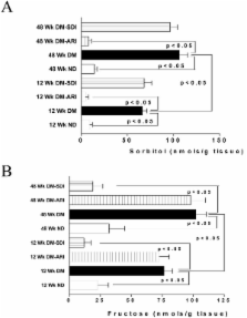
          We investigated the role of polyol pathway enzymes aldose reductase (AR) and sorbitol dehydrogenase (SDH) in mediating injury due to ischemia-reperfusion (IR) in Type 2 diabetic BBZ rat hearts. Specifically, we investigated, (a) changes in glucose flux via cardiac AR and SDH as a function of diabetes duration, (b) ischemic injury and function after IR, (c) the effect of inhibition of AR or SDH on ischemic injury and function. Hearts isolated from BBZ rats, after 12 weeks or 48 weeks diabetes duration, and their non-diabetic littermates, were subjected to IR protocol. Myocardial function, substrate flux via AR and SDH, and tissue lactate:pyruvate (L/P) ratio (a measure of cytosolic NADH/NAD +), and lactate dehydrogenase (LDH) release (a marker of IR injury) were measured. Zopolrestat, and CP-470,711 were used to inhibit AR and SDH, respectively. Myocardial sorbitol and fructose content, and associated changes in L/P ratios were significantly higher in BBZ rats compared to non-diabetics, and increased with disease duration. Induction of IR resulted in increased ischemic injury, reduced ATP levels, increases in L/P ratio, and poor cardiac function in BBZ rat hearts, while inhibition of AR or SDH attenuated these changes and protected hearts from IR injury. These data indicate that AR and SDH are key modulators of myocardial IR injury in BBZ rat hearts and that inhibition of polyol pathway could in principle be used as a therapeutic adjunct for protection of ischemic myocardium in Type 2 diabetic patients.

          Related collections

          Most cited references55

          • Record: found
          • Abstract: found
          • Article: found
          Is Open Access

          Oxidative stress and the use of antioxidants in diabetes: Linking basic science to clinical practice

          Cardiovascular complications, characterized by endothelial dysfunction and accelerated atherosclerosis, are the leading cause of morbidity and mortality associated with diabetes. There is growing evidence that excess generation of highly reactive free radicals, largely due to hyperglycemia, causes oxidative stress, which further exacerbates the development and progression of diabetes and its complications. Overproduction and/or insufficient removal of these free radicals result in vascular dysfunction, damage to cellular proteins, membrane lipids and nucleic acids. Despite overwhelming evidence on the damaging consequences of oxidative stress and its role in experimental diabetes, large scale clinical trials with classic antioxidants failed to demonstrate any benefit for diabetic patients. As our understanding of the mechanisms of free radical generation evolves, it is becoming clear that rather than merely scavenging reactive radicals, a more comprehensive approach aimed at preventing the generation of these reactive species as well as scavenging may prove more beneficial. Therefore, new strategies with classic as well as new antioxidants should be implemented in the treatment of diabetes.
            Bookmark
            • Record: found
            • Abstract: found
            • Article: not found

            Myocardial cell death in human diabetes.

            The renin-angiotensin system is upregulated with diabetes, and this may contribute to the development of a dilated myopathy. Angiotensin II (Ang II) locally may lead to oxidative damage, activating cardiac cell death. Moreover, diabetes and hypertension could synergistically impair myocardial structure and function. Therefore, apoptosis and necrosis were measured in ventricular myocardial biopsies obtained from diabetic and diabetic-hypertensive patients. Accumulation of a marker of oxidative stress, nitrotyrosine, and Ang II labeling were evaluated quantitatively. The diabetic heart showed cardiac hypertrophy, cavitary dilation, and depressed ventricular performance. These alterations were more severe with diabetes and hypertension. Diabetes was characterized by an 85-fold, 61-fold, and 26-fold increase in apoptosis of myocytes, endothelial cells, and fibroblasts, respectively. Apoptosis in cardiac cells did not increase additionally with diabetes and hypertension. Diabetes increased necrosis by 4-fold in myocytes, 9-fold in endothelial cells, and 6-fold in fibroblasts. However, diabetes and hypertension increased necrosis by 7-fold in myocytes and 18-fold in endothelial cells. Similarly, Ang II labeling in myocytes and endothelial cells increased more with diabetes and hypertension than with diabetes alone. Nitrotyrosine localization in cardiac cells followed a comparable pattern. In spite of the difference in the number of nitrotyrosine-positive cells with diabetes and with diabetes and hypertension, apoptosis and necrosis of myocytes, endothelial cells, and fibroblasts were detected only in cells containing this modified amino acid. In conclusion, local increases in Ang II with diabetes and with diabetes and hypertension may enhance oxidative damage, activating cardiac cell apoptosis and necrosis.
              Bookmark
              • Record: found
              • Abstract: found
              • Article: not found

              Hyperglycemic pseudohypoxia and diabetic complications.

              Vasodilation and increased blood flow are characteristic early vascular responses to acute hyperglycemia and tissue hypoxia. In hypoxic tissues these vascular changes are linked to metabolic imbalances associated with impaired oxidation of NADH to NAD+ and the resulting increased ratio of NADH/NAD+. In hyperglycemic tissues these vascular changes also are linked to an increased ratio of NADH/NAD+, in this case because of an increased rate of reduction of NAD+ to NADH. Several lines of evidence support the likelihood that the increased cytosolic ratio of free NADH/NAD+ caused by hyperglycemia, referred to as pseudohypoxia because tissue partial pressure oxygen is normal, is a characteristic feature of poorly controlled diabetes that mimics the effects of true hypoxia on vascular and neural function and plays an important role in the pathogenesis of diabetic complications. These effects of hypoxia and hyperglycemia-induced pseudohypoxia on vascular and neural function are mediated by a branching cascade of imbalances in lipid metabolism, increased production of superoxide anion, and possibly increased nitric oxide formation.
                Bookmark

                Author and article information

                Journal
                Cardiovasc Diabetol
                Cardiovascular Diabetology
                BioMed Central
                1475-2840
                2008
                28 October 2008
                : 7
                : 33
                Affiliations
                [1 ]Division of Surgical Science, Department of Surgery, Columbia University College of Physicians and Surgeons, New York, NY 10032, USA
                [2 ]Pfizer Global Research, Groton, CT, USA
                [3 ]Biomedical Research Models Inc, Worcester, MA, USA
                Article
                1475-2840-7-33
                10.1186/1475-2840-7-33
                2584021
                18957123
                e9e19134-5b3d-4dc6-9b18-b1f56d8e4820
                Copyright © 2008 Li et al; licensee BioMed Central Ltd.

                This is an Open Access article distributed under the terms of the Creative Commons Attribution License ( http://creativecommons.org/licenses/by/2.0), which permits unrestricted use, distribution, and reproduction in any medium, provided the original work is properly cited.

                History
                : 14 August 2008
                : 28 October 2008
                Categories
                Original Investigation

                Endocrinology & Diabetes
                Endocrinology & Diabetes

                Comments

                Comment on this article