46
views
0
recommends
+1 Recommend
0 collections
    0
    shares
      • Record: found
      • Abstract: found
      • Article: found
      Is Open Access

      Acute Zonal Occult Outer Retinopathy in Japanese Patients: Clinical Features, Visual Function, and Factors Affecting Visual Function

      research-article

      Read this article at

      Bookmark
          There is no author summary for this article yet. Authors can add summaries to their articles on ScienceOpen to make them more accessible to a non-specialist audience.

          Abstract

          Purpose

          To evaluate the clinical features and investigate their relationship with visual function in Japanese patients with acute zonal occult outer retinopathy (AZOOR).

          Methods

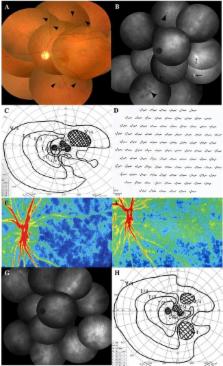
          Fifty-two eyes of 38 Japanese AZOOR patients (31 female and 7 male patients; mean age at first visit, 35.0 years; median follow-up duration, 31 months) were retrospectively collected: 31 untreated eyes with good visual acuity and 21 systemic corticosteroid-treated eyes with progressive visual acuity loss. Variables affecting the logMAR values of best-corrected visual acuity (BCVA) and the mean deviation (MD) on Humphrey perimetry at initial and final visits were examined using multiple stepwise linear regression analysis.

          Results

          In untreated eyes, the mean MD at the final visit was significantly higher than that at the initial visit ( P = 0.00002). In corticosteroid-treated eyes, the logMAR BCVA and MD at the final visit were significantly better than the initial values ( P = 0.007 and P = 0.02, respectively). The final logMAR BCVA was 0.0 or less in 85% of patients. Variables affecting initial visual function were moderate anterior vitreous cells, myopia severity, and a-wave amplitudes on electroretinography; factors affecting final visual function were the initial MD values, female sex, moderate anterior vitreous cells, and retinal atrophy.

          Conclusions

          Our data indicated that visual functions in enrolled patients significantly improved spontaneously or after systemic corticosteroids therapy, suggesting that Japanese patients with AZOOR have good visual outcomes during the follow-up period of this study. Furthermore, initial visual field defects, gender, anterior vitreous cells, and retinal atrophy affected final visual functions in these patients.

          Related collections

          Most cited references21

          • Record: found
          • Abstract: found
          • Article: not found

          Acute zonal occult outer retinopathy: a long-term follow-up study.

          To report the long-term follow-up of patients with acute zonal occult outer retinopathy (AZOOR). Observational consecutive case series. Prospective and retrospective review of medical records of patients with AZOOR. Fifty-one patients (37 women and 14 men) with a median age of 33 years (mean, age 36 years; range, 13-63 years) were followed for a median of 96 months (mean 100 months; range, 36-420 months). At presentation, AZOOR was present in one eye of 31 patients (61%) and both eyes in 20 patients (39%). All patients presented with an acute loss of one or more zones of visual field, and 45 (88%) patients presented with photopsia. Corrected visual acuity was 20/40 or better in 68 (76%) of 90 affected eyes. Funduscopic examination was normal in 82 eyes and revealed signs of AZOOR in 8 eyes. Electroretinographic amplitudes were depressed in all affected eyes. The median delay in diagnosis of AZOOR was 17 months. During follow-up, AZOOR developed in 19 fellow eyes. At final follow-up AZOOR was present in one eye of 12 (24%) patients and both eyes of 39 (76%) patients. Sixteen patients had 23 recurrences of AZOOR. Visual field loss stabilized within 6 months in 37 patients (72%), progressed stepwise in 2 patients (4%), and partly improved in 12 patients (24%). Fourteen patients (28%) had autoimmune diseases, including Hashimoto's thyroiditis in 6 patients and relapsing transverse myelopathy in 4 patients. At last follow-up all patients had residual visual field defects. Final visual acuity was 20/40 or better in 61 (68%) affected eyes. Nine patients (18%) were legally blind. The fundi of 90 affected eyes revealed no changes of AZOOR in 47 eyes (52%) and changes in the pigment epithelium and retina caused by AZOOR in 43 eyes (48%). Visual loss in AZOOR is characterized by one or more episodes of acute dysfunction, and in some cases, death of retinal receptor cells in one or more zones of one or both eyes. Central vision is often spared, but recovery of visual field occurs infrequently. The etiology of AZOOR is unknown. Electroretinography is essential for early diagnosis. The value of treatment is uncertain.
            Bookmark
            • Record: found
            • Abstract: found
            • Article: not found

            Photoreceptor outer segment abnormalities as a cause of blind spot enlargement in acute zonal occult outer retinopathy-complex diseases.

            To investigate the correlation between visual field (VF) defects in diseases of the acute zonal occult outer retinopathy (AZOOR)-complex and their spectral-domain optical coherence tomographic (OCT) findings. Observational case series. Patients with AZOOR, multiple evanescent white dot syndrome (MEWDS), and multifocal choroiditis and panuveitis (MCP) examined in a private practice retinal referral center had threshold VF testing and spectral-domain OCT examination performed using a device capable of obtaining a block of 128 B-scans in a 6 x 6-mm region centered on the optic nerve and macula. The areas of defects in the boundary between the inner segments (IS) and the outer segments (OS) of the photoreceptors, termed the IS/OS boundary, were compared with the VF defects measured. There were 18 evaluable eyes among one patient with MEWDS, two with AZOOR, and seven with MCP. In the 14 eyes with blind spot enlargement [corrected] corresponding IS/OS boundary defects were found in the [corrected] peripapillary region, while no IS/OS boundary defects were found in the four [corrected] eyes without blind spot enlargement. IS/OS boundary defects were seen over chorioretinal scars and areas of neovascularization and no widespread defects were seen [corrected] elsewhere in the fundus. The IS/OS boundary defects showed improvement, as did the blind spot enlargement, spontaneously in the patient with MEWDS and after treatment with immunosuppression in the patients with AZOOR. The spectral-domain OCT finding of IS/OS boundary defects, implicating photoreceptor OS perturbation, appears to explain the blind spot enlargement in patients with AZOOR-complex diseases. These defects are not necessarily permanent.
              Bookmark
              • Record: found
              • Abstract: found
              • Article: not found

              Acute zonal occult outer retinopathy. Donders Lecture: The Netherlands Ophthalmological Society, Maastricht, Holland, June 19, 1992.

              J Gass (1993)
              This report describes 13 patients, predominantly young women, with a syndrome characterized by rapid loss of one or more large zones of outer retinal function, photopsia, minimal funduscopic changes, and electroretinographic abnormalities affecting one or both eyes. All patients on follow-up examination had persistent visual field defects, and most had chronic photopsia and zones of pigment epithelial atrophy. Evidence is presented that these patients probably represent part of the spectrum of a single disorder that includes the multiple evanescent white-dot syndrome (MEWDS), acute idiopathic blind-spot-enlargement syndrome (AIBSES), acute macular neuroretinopathy (AMN), and the pseudo-presumed ocular histoplasmosis syndrome (P-POHS). The medical records of these 13 patients and 2 additional patients, who developed, in addition to the features of this syndrome, funduscopic changes typical of MEWDS, AMN, and P-POHS, were reviewed and follow-up obtained. These patients had extensive unrewarding medical and neurological investigations because of suspected diagnoses, including central nervous system disorders, cancer-associated retinopathy, retinal vasculitis, diffuse unilateral subacute neuroretinitis, and tapetoretinal degenerations. Although most patients retained good visual acuity, all had permanent visual field loss that in some cases was severe. The cause of the disorder was not determined. No effective treatment was found. Acute visual loss and photopsia in these patients is probably caused by damage to large zones of the outer retina that appears unaffected ophthalmoscopically. Electroretinography is important in early diagnosis. Future investigations probably will reveal further evidence linking this disorder to MEWDS, AIBSES, AMN, and P-POHS.
                Bookmark

                Author and article information

                Contributors
                Role: Academic Editor
                Journal
                PLoS One
                PLoS ONE
                plos
                plosone
                PLoS ONE
                Public Library of Science (San Francisco, CA USA )
                1932-6203
                28 April 2015
                2015
                : 10
                : 4
                : e0125133
                Affiliations
                [1 ]Department of Ophthalmology, Hokkaido University Graduate School of Medicine, Sapporo, Hokkaido, Japan
                [2 ]Department of Ocular Circulation and Metabolism, Hokkaido University Graduate School of Medicine, Sapporo, Hokkaido, Japan
                Saitama Medical University, JAPAN
                Author notes

                Competing Interests: The authors have declared that no competing interests exist.

                Conceived and designed the experiments: SS WS MS SI. Performed the experiments: SS WS YH SM K. Noda K. Namba. Analyzed the data: SS WS MS YH SM K. Noda K. Namba SI. Wrote the paper: SS WS MS YH K. Noda SI. Statistical analysis: MS. Critical revision of the manuscript: WS SM K. Noda K. Namba SI.

                Article
                PONE-D-14-46510
                10.1371/journal.pone.0125133
                4412707
                25919689
                eab5f077-799d-44b9-9a27-f539a44b658b
                Copyright @ 2015

                This is an open access article distributed under the terms of the Creative Commons Attribution License, which permits unrestricted use, distribution, and reproduction in any medium, provided the original author and source are credited

                History
                : 20 October 2014
                : 11 March 2015
                Page count
                Figures: 6, Tables: 4, Pages: 16
                Funding
                The authors have no support or funding to report.
                Categories
                Research Article
                Custom metadata
                All relevant data are within the paper.

                Uncategorized
                Uncategorized

                Comments

                Comment on this article