1
views
0
recommends
+1 Recommend
0 collections
    0
    shares
      • Record: found
      • Abstract: found
      • Article: found
      Is Open Access

      Probing key elements of teixobactin–lipid II interactions in membranes†

      research-article

      Read this article at

      Bookmark
          There is no author summary for this article yet. Authors can add summaries to their articles on ScienceOpen to make them more accessible to a non-specialist audience.

          Abstract

          Two binding poses of the teixobactin–lipid II complex were captured with MD simulations at the membrane surface.

          Abstract

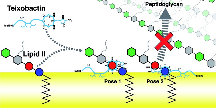
          Teixobactin (Txb) is a recently discovered antibiotic against Gram-positive bacteria that induces no detectable resistance. The bactericidal mechanism is believed to be the inhibition of cell wall biosynthesis by Txb binding to lipid II and lipid III. Txb binding specificity likely arises from targeting of the shared lipid component, the pyrophosphate moiety. Despite synthesis and functional assessment of numerous chemical analogs of Txb, and consequent identification of the Txb pharmacophore, the detailed structural information of Txb–substrate binding is still lacking. Here, we use molecular modeling and microsecond-scale molecular dynamics simulations to capture the formation of Txb–lipid II complexes at a membrane surface. Two dominant binding conformations were observed, both showing characteristic lipid II phosphate binding by the Txb backbone amides near the C-terminal cyclodepsipeptide ( d-Thr8–Ile11) ring. Additionally, binding by Txb also involved the side chain hydroxyl group of Ser7, as well as a secondary phosphate binding provided by the side chain of l- allo-enduracididine. Interestingly, those conformations differ by swapping two groups of hydrogen bond donors that coordinate the two phosphate moieties of lipid II, resulting in opposite orientations of lipid II binding. In addition, residues d- allo-Ile5 and Ile6 serve as the membrane anchors in both Txb conformations, regardless of the detailed phosphate binding interactions near the cyclodepsipeptide ring. The role of hydrophobic residues in Txb activity is primarily for its membrane insertion, and subsidiarily to provide non-polar interactions with the lipid II tail. Based on the Txb–lipid II interactions captured in their complexes, as well as their partitioning depths into the membrane, we propose that the bactericidal mechanism of Txb is to arrest cell wall synthesis by selectively inhibiting the transglycosylation of peptidoglycan, while possibly leaving the transpeptidation step unaffected. The observed “pyrophosphate caging” mechanism of lipid II inhibition appears to be similar to some lantibiotics, but different from that of vancomycin or bacitracin.

          Related collections

          Most cited references3

          • Record: found
          • Abstract: found
          • Article: not found

          CHARMM additive all-atom force field for carbohydrate derivatives and its utility in polysaccharide and carbohydrate-protein modeling.

          Monosaccharide derivatives such as xylose, fucose, N-acetylglucosamine (GlcNAc), N-acetylgalactosamine (GlaNAc), glucuronic acid, iduronic acid, and N-acetylneuraminic acid (Neu5Ac) are important components of eukaryotic glycans. The present work details development of force-field parameters for these monosaccharides and their covalent connections to proteins via O-linkages to serine or threonine sidechains and via N-linkages to asparagine sidechains. The force field development protocol was designed to explicitly yield parameters that are compatible with the existing CHARMM additive force field for proteins, nucleic acids, lipids, carbohydrates, and small molecules. Therefore, when combined with previously developed parameters for pyranose and furanose monosaccharides, for glycosidic linkages between monosaccharides, and for proteins, the present set of parameters enables the molecular simulation of a wide variety of biologically-important molecules such as complex carbohydrates and glycoproteins. Parametrization included fitting to quantum mechanical (QM) geometries and conformational energies of model compounds, as well as to QM pair interaction energies and distances of model compounds with water. Parameters were validated in the context of crystals of relevant monosaccharides, as well NMR and/or x-ray crystallographic data on larger systems including oligomeric hyaluronan, sialyl Lewis X, O- and N-linked glycopeptides, and a lectin:sucrose complex. As the validated parameters are an extension of the CHARMM all-atom additive biomolecular force field, they further broaden the types of heterogeneous systems accessible with a consistently-developed force-field model.
            Bookmark
            • Record: found
            • Abstract: not found
            • Article: not found

            Hydrogen bonding in globular proteins

              Bookmark
              • Record: found
              • Abstract: found
              • Article: not found

              The nisin-lipid II complex reveals a pyrophosphate cage that provides a blueprint for novel antibiotics.

              The emerging antibiotics-resistance problem has underlined the urgent need for novel antimicrobial agents. Lantibiotics (lanthionine-containing antibiotics) are promising candidates to alleviate this problem. Nisin, a member of this family, has a unique pore-forming activity against bacteria. It binds to lipid II, the essential precursor of cell wall synthesis. As a result, the membrane permeabilization activity of nisin is increased by three orders of magnitude. Here we report the solution structure of the complex of nisin and lipid II. The structure shows a novel lipid II-binding motif in which the pyrophosphate moiety of lipid II is primarily coordinated by the N-terminal backbone amides of nisin via intermolecular hydrogen bonds. This cage structure provides a rationale for the conservation of the lanthionine rings among several lipid II-binding lantibiotics. The structure of the pyrophosphate cage offers a template for structure-based design of novel antibiotics.
                Bookmark

                Author and article information

                Journal
                Chem Sci
                Chem Sci
                Chemical Science
                Royal Society of Chemistry
                2041-6520
                2041-6539
                20 July 2018
                14 September 2018
                : 9
                : 34
                : 6997-7008
                Affiliations
                [a ] Department of Biochemistry , Center for Biophysics and Quantitative Biology , Beckman Institute for Advanced Science and Technology , University of Illinois at Urbana-Champaign , Urbana , Illinois 61801 , USA . Email: pwen2@ 123456illinois.edu ; Email: emad@ 123456life.illinois.edu
                [b ] Department of Nanobiology , Center for Biological and Engineering Sciences , Sandia National Laboratories , Albuquerque , NM 87185 , USA . Email: slrempe@ 123456sandia.gov
                Author notes

                ‡Present address: Department of Physics, University of Vermont, Burlington, VT 05405, U.S.A. Email: Email: jvanegas@ 123456uvm.edu

                Author information
                http://orcid.org/0000-0002-6049-6904
                http://orcid.org/0000-0003-1623-2108
                http://orcid.org/0000-0001-8434-1010
                Article
                c8sc02616e
                10.1039/c8sc02616e
                6124899
                30210775
                eab81409-6ee7-4ac8-b902-6a815aefb613
                This journal is © The Royal Society of Chemistry 2018

                This article is freely available. This article is licensed under a Creative Commons Attribution 3.0 Unported Licence (CC BY 3.0)

                History
                : 13 June 2018
                : 19 July 2018
                Categories
                Chemistry

                Notes

                †Electronic supplementary information (ESI) available: Computational methods and supplementary figures. See DOI: 10.1039/c8sc02616e


                Comments

                Comment on this article