28
views
0
recommends
+1 Recommend
0 collections
    0
    shares
      • Record: found
      • Abstract: found
      • Article: found
      Is Open Access

      Cardiovascular magnetic resonance parameters associated with early transplant-free survival in children with small left hearts following conversion from a univentricular to biventricular circulation

      research-article

      Read this article at

      Bookmark
          There is no author summary for this article yet. Authors can add summaries to their articles on ScienceOpen to make them more accessible to a non-specialist audience.

          Abstract

          Background

          We sought to identify cardiovascular magnetic resonance (CMR) parameters associated with successful univentricular to biventricular conversion in patients with small left hearts.

          Methods

          Patients with small left heart structures and a univentricular circulation who underwent CMR prior to biventricular conversion were retrospectively identified and divided into 2 anatomic groups: 1) borderline hypoplastic left heart structures (BHLHS), and 2) right-dominant atrioventricular canal (RDAVC). The primary outcome variable was transplant-free survival with a biventricular circulation.

          Results

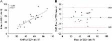
          In the BHLHS group (n = 22), 16 patients (73%) survived with a biventricular circulation over a median follow-up of 40 months (4–84). Survival was associated with a larger CMR left ventricular (LV) end-diastolic volume (EDV) (p = 0.001), higher LV-to-right ventricle (RV) stroke volume ratio (p < 0.001), and higher mitral-to-tricuspid inflow ratio (p = 0.04). For predicting biventricular survival, the addition of CMR threshold values to echocardiographic LV EDV improved sensitivity from 75% to 93% while maintaining specificity at 100%. In the RDAVC group (n = 10), 9 patients (90%) survived with a biventricular circulation over a median follow-up of 29 months (3–51). The minimum CMR values were a LV EDV of 22 ml/m 2 and a LV-to-RV stroke volume ratio of 0.19.

          Conclusions

          In BHLHS patients, a larger LV EDV, LV-to-RV stroke volume ratio, and mitral-to-tricuspid inflow ratio were associated with successful biventricular conversion. The addition of CMR parameters to echocardiographic measurements improved the sensitivity for predicting successful conversion. In RDAVC patients, the high success rate precluded discriminant analysis, but a range of CMR parameters permitting biventricular conversion were identified.

          Related collections

          Most cited references27

          • Record: found
          • Abstract: not found
          • Article: not found

          Guidelines and standards for performance of a pediatric echocardiogram: a report from the Task Force of the Pediatric Council of the American Society of Echocardiography.

            Bookmark
            • Record: found
            • Abstract: found
            • Article: not found

            Mitral valve and tricuspid valve blood flow: accurate quantification with 3D velocity-encoded MR imaging with retrospective valve tracking.

            To validate flow assessment performed with three-dimensional (3D) three-directional velocity-encoded (VE) magnetic resonance (MR) imaging with retrospective valve tracking and to compare this modality with conventional two-dimensional (2D) one-directional VE MR imaging in healthy subjects and patients with regurgitation. Patients and volunteers gave informed consent, and local medical ethics committee approval was obtained. Patient data were selected retrospectively and randomly from a database of MR studies obtained between July 2006 and July 2007. The 3D three-directional VE MR images were first validated in vitro and compared with 2D one-directional VE MR images. Mitral valve (MV) and tricuspid valve (TV) flow were assessed in 10 volunteers without valve insufficiency and 20 patients with valve insufficiency, with aortic systolic stroke volume (ASSV) as the reference standard. Phantom validation showed less than 5% error for both techniques. In volunteers, 3D three-directional VE MR images showed no bias for MV or TV flow when compared with ASSV, whereas 2D one-directional VE MR images showed significant bias for MV flow (15% overestimation, P < .01). TV flow showed 25% overestimation; however, this was insignificant because of the high standard deviation. Correlation with ASSV was strong for 3D three-directional VE MR imaging (r = 0.96, P < .01 for MV flow; r = 0.88, P < .01 for TV flow) and between MV and TV flow (r = 0.91, P < .01); however, correlation was weaker for 2D one-directional VE MR imaging (r = 0.80, P < .01 for MV flow; r = 0.22, P = .55 for TV flow) and between MV flow and TV flow (r = 0.34, P = .34). In patients (mean regurgitation fractions of 13% and 10% for MV flow and TV flow, respectively), correlation between MV flow and TV flow for 3D three-directional VE MR imaging was strong (r = 0.97, P < .01). Use of 3D three-directional VE MR imaging enables accurate MV and TV flow quantification, even in patients with valve regurgitation. RSNA, 2008
              Bookmark
              • Record: found
              • Abstract: found
              • Article: found
              Is Open Access

              Normal right- and left ventricular volumes and myocardial mass in children measured by steady state free precession cardiovascular magnetic resonance

              Background Quantification of ventricular volume by Steady State Free Precession (SSFP) cardiovascular magnetic resonance is accurate and reproducible. Normal values exist for adults, but are lacking for children. We sought to establish normal values for left and right ventricular volumes, mass and function in healthy children by using SSFP. Methods and results Fifty children (27 females, 23 males) without cardiovascular disease were evaluated. Median age was 11 years (range 7 months – 18 years), weight 35 kg (range 7–77 kg), height 146 cm (range 66–181 cm). Thirty-six examinations were performed with breath holding, 14 in freely breathing sedated children. Ventricular volumes and mass were measured in the end systolic and end diastolic phase on SSFP cine images acquired in a short axis plane as a stack of 12 contiguous slices covering full length of both ventricles. Regression analysis showed an exponential relationship between body surface area (BSA) and ventricular volumes and mass (normal value = a*BSAb ). Normative curves for males and females are presented in relation to BSA for the enddiastolic volume, endsystolic volume and mass of both ventricles. Intra- and interobserver variability of the measurements was within the limits of 2% and 7% respectively, except for right ventricular mass (10%). Conclusion The exponential equation for calculation of normal values for each ventricular parameter and graphical display of normative curves for data acquired in healthy children by SSFP cardiovascular magnetic resonance are provided.
                Bookmark

                Author and article information

                Contributors
                puja.banka@cardio.chboston.org
                barbara.schaetzle@hotmail.com
                rukmini.komarlu@cardio.chboston.org
                sitaram.emani@cardio.chboston.org
                tal.geva@cardio.chboston.org
                andrew.powell@cardio.chboston.org
                Journal
                J Cardiovasc Magn Reson
                J Cardiovasc Magn Reson
                Journal of Cardiovascular Magnetic Resonance
                BioMed Central (London )
                1097-6647
                1532-429X
                7 October 2014
                7 October 2014
                2014
                : 16
                : 1
                : 73
                Affiliations
                [ ]Department of Cardiology, Boston Children’s Hospital, 300 Longwood Ave, Boston, MA 02115 USA
                [ ]Department of Cardiac Surgery, Boston Children’s Hospital, 300 Longwood Ave, Boston, MA 02115 USA
                [ ]Department of Pediatrics, Harvard Medical School, Boston, MA USA
                [ ]Department of Surgery, Harvard Medical School, Boston, MA USA
                [ ]Current address: Kantonsspital Winterthur, Winterthur, Switzerland
                [ ]Current address: Department of Pediatric Cardiology, Cleveland Clinic, Cleveland, OH USA
                Article
                73
                10.1186/s12968-014-0073-1
                4189673
                25314952
                eaffbb57-d56b-4536-b01e-15681a961bca
                © Banka et al.; licensee BioMed Central Ltd. 2014

                This is an Open Access article distributed under the terms of the Creative Commons Attribution License ( http://creativecommons.org/licenses/by/4.0), which permits unrestricted use, distribution, and reproduction in any medium, provided the original work is properly credited. The Creative Commons Public Domain Dedication waiver ( http://creativecommons.org/publicdomain/zero/1.0/) applies to the data made available in this article, unless otherwise stated.

                History
                : 8 April 2014
                : 27 August 2014
                Categories
                Research
                Custom metadata
                © The Author(s) 2014

                Cardiovascular Medicine
                hypoplastic left heart syndrome,atrioventricular canal,cardiovascular magnetic resonance,pediatrics,congenital heart disease,surgery,biventricular repair

                Comments

                Comment on this article