17
views
0
recommends
+1 Recommend
0 collections
    0
    shares
      • Record: found
      • Abstract: found
      • Article: found
      Is Open Access

      Anesthetic Isoflurane Increases Phosphorylated Tau Levels Mediated by Caspase Activation and Aβ Generation

      research-article

      Read this article at

      Bookmark
          There is no author summary for this article yet. Authors can add summaries to their articles on ScienceOpen to make them more accessible to a non-specialist audience.

          Abstract

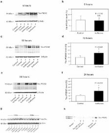
          Anesthetic isoflurane has been shown to promote Alzheimer’s disease (AD) neuropathogenesis by inducing caspase activation and accumulation of β-amyloid (Aβ). Phosphorylation of tau protein is another important feature of AD neuropathogenesis. However, the effects of isoflurane on phosphorylated tau levels remain largely to be determined. We therefore set out to determine whether isoflurane can increase phosphorylated tau levels. 5 to 8 month-old wild-type and AD transgenic mice [B6.Cg-Tg (APPswe, PSEN1dE9)85Dbo/J] were treated with 1.4% isoflurane for two hours. The mice brain tissues were harvested at six, 12 and 24 hours after the anesthesia. For the in vitro studies, primary neurons from wild-type and the AD transgenic mice were exposed to 2% isoflurane for six hours, and were harvested at the end of anesthesia. The harvested brain tissues and neurons were subjected to Western blot analysis by which the levels of phosphorylated tau protein at Serine 262 (Tau-PS262) were determined. Here we show that the isoflurane anesthesia increased Tau-PS262 levels in brain tissues and primary neurons from the wild-type and AD transgenic mice. Moreover, the isoflurane anesthesia may induce a greater increase in Tau-PS262 levels in primary neurons and brain tissues from the AD transgenic mice. Finally, caspase activation inhibitor Z-VAD and Aβ generation inhibitor L-685,458 attenuated the isoflurane-induced increases in Tau-PS262 levels. In conclusion, clinically relevant isoflurane anesthesia increases phosphorylated tau levels, which may result from the isoflurane-induced caspase activation and Aβ generation. These findings will promote more studies to determine the effects of anesthetics on tau phosphorylation.

          Related collections

          Most cited references39

          • Record: found
          • Abstract: found
          • Article: not found

          Tau protein isoforms, phosphorylation and role in neurodegenerative disorders.

          Tau proteins belong to the family of microtubule-associated proteins. They are mainly expressed in neurons where they play an important role in the assembly of tubulin monomers into microtubules to constitute the neuronal microtubules network. Microtubules are involved in maintaining the cell shape and serve as tracks for axonal transport. Tau proteins also establish some links between microtubules and other cytoskeletal elements or proteins. Tau proteins are translated from a single gene located on chromosome 17. Their expression is developmentally regulated by an alternative splicing mechanism and six different isoforms exist in the human adult brain. Tau proteins are the major constituents of intraneuronal and glial fibrillar lesions described in Alzheimer's disease and numerous neurodegenerative disorders referred to as 'tauopathies'. Molecular analysis has revealed that an abnormal phosphorylation might be one of the important events in the process leading to their aggregation. Moreover, a specific set of pathological tau proteins exhibiting a typical biochemical pattern, and a different regional and laminar distribution could characterize each of these disorders. Finally, a direct correlation has been established between the progressive involvement of the neocortical areas and the increasing severity of dementia, suggesting that pathological tau proteins are reliable marker of the neurodegenerative process. The recent discovery of tau gene mutations in frontotemporal dementia with parkinsonism linked to chromosome 17 has reinforced the predominant role attributed to tau proteins in the pathogenesis of neurodegenerative disorders, and underlined the fact that distinct sets of tau isoforms expressed in different neuronal populations could lead to different pathologies.
            Bookmark
            • Record: found
            • Abstract: found
            • Article: not found

            Linking Abeta and tau in late-onset Alzheimer's disease: a dual pathway hypothesis.

            Alzheimer's disease is characterized by abnormal elevation of Abeta peptide and abnormal hyperphosphorylation of the tau protein. The "amyloid hypothesis," which is based on molecular defects observed in autosomal-dominant early-onset Alzheimer's disease (EOAD), suggests a serial model of causality, whereby elevation of Abeta drives other disease features including tau hyperphosphorylation. Here, we review recent evidence from drug trials, genetic studies, and experimental work in animal models that suggests that an alternative model might exist in late-onset AD (LOAD), the complex and more common form of the disease. Specifically, we hypothesize a "dual pathway" model of causality, whereby Abeta and tau can be linked by separate mechanisms driven by a common upstream driver. This model may account for the results of recent drug trials and, if confirmed, may guide future drug development.
              Bookmark
              • Record: found
              • Abstract: found
              • Article: not found

              Specific tau phosphorylation sites correlate with severity of neuronal cytopathology in Alzheimer's disease.

              Microtubule associated protein tau is abnormally phosphorylated in Alzheimer's disease (AD) and aggregates as paired helical filaments (PHFs) in neurofibrillary tangles (NFTs). We show here that the pattern of tau phosphorylation correlates with the loss of neuronal integrity. Studies using 11 phosphorylation dependent tau antibodies and a panel of AD cases of varying severity were evaluated in terms of three stages of neurofibrillary tangle development: (1) pre-neurofibrillary tangle, (2) intra-, and (3) extra-neuronal neurofibrillary tangles. The pretangle state, in which neurons display nonfibrillar, punctate regions in the cytoplasm, sound dendrites, somas, and nuclei, was observed especially with phospho-tau antibodies TG3 (pT231), pS262, and pT153. Intraneuronal neurofibrillary tangles are homogenously stained with fibrillar tau structures, which were most prominently stained with pT175/181, 12E8 (pS262/pS356), pS422, pS46, pS214 antibodies. Extracellular NFTs, which contain substantial filamentous tau, are most prominently stained with AT8 (pS199/pS202/pT205), AT100 (pT212/pS214), and PHF-1 (pS396/pS404) antibodies, which also stain intracellular NFT. The sequence of early tau phosphorylation suggests that there are events prior to filament formation that are specific to particular phosphorylated tau epitopes, leading to conformational changes and cytopathological alterations.
                Bookmark

                Author and article information

                Contributors
                Role: Editor
                Journal
                PLoS One
                PLoS ONE
                plos
                plosone
                PLoS ONE
                Public Library of Science (San Francisco, USA )
                1932-6203
                2012
                20 June 2012
                : 7
                : 6
                : e39386
                Affiliations
                [1 ]Geriatric Anesthesia Research Unit, Department of Anesthesia, Critical Care and Pain Medicine, Massachusetts General Hospital and Harvard Medical School, Charlestown, Massachusetts, United States of America
                [2 ]Department of Forensic Pathology, Faculty of Forensic Medicine, China Medical University, Shenyang, People’s Republic of China
                University of Nebraska Medical Center, United States of America
                Author notes

                Conceived and designed the experiments: YD XW Z. Xu YZ. Performed the experiments: YD XW. Analyzed the data: Z. Xu YZ Z. Xie. Wrote the paper: Z. Xie.

                Article
                PONE-D-12-09437
                10.1371/journal.pone.0039386
                3379981
                22745746
                ec9b5159-375b-4c39-a00c-fdf90694aabe
                Dong et al. This is an open-access article distributed under the terms of the Creative Commons Attribution License, which permits unrestricted use, distribution, and reproduction in any medium, provided the original author and source are credited.
                History
                : 31 March 2012
                : 23 May 2012
                Page count
                Pages: 12
                Categories
                Research Article
                Biology
                Biochemistry
                Cytochemistry
                Histochemistry
                Developmental Biology
                Molecular Development
                Cytokines
                Immunology
                Immunologic Techniques
                Immunohistochemical Analysis
                Model Organisms
                Animal Models
                Mouse
                Molecular Cell Biology
                Neuroscience
                Cellular Neuroscience
                Medicine
                Anesthesiology
                General Anesthesia
                Neurology
                Dementia
                Alzheimer Disease
                Neurodegenerative Diseases

                Uncategorized
                Uncategorized

                Comments

                Comment on this article