5
views
0
recommends
+1 Recommend
0 collections
    0
    shares
      • Record: found
      • Abstract: found
      • Article: found
      Is Open Access

      Valvular imaging in the era of feature‐tracking: A slice‐following cardiac MR sequence to measure mitral flow

      research-article

      Read this article at

      Bookmark
          There is no author summary for this article yet. Authors can add summaries to their articles on ScienceOpen to make them more accessible to a non-specialist audience.

          Abstract

          Background

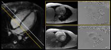
          In mitral valve dysfunction, noninvasive measurement of transmitral blood flow is an important clinical examination. Flow imaging of the mitral valve, however, is challenging, since it moves in and out of the image plane during the cardiac cycle.

          Purpose

          To more accurately measure mitral flow, a slice‐following MRI phase contrast sequence is proposed. This study aimed to implement such a sequence, validate its slice‐following functionality in a phantom and healthy subjects, and test its feasibility in patients with mitral valve dysfunction.

          Study Type

          Prospective.

          Phantom and Subjects

          The slice‐following functionality was validated in a cone‐shaped phantom by measuring the depicted slice radius. Sixteen healthy subjects and 10 mitral valve dysfunction patients were enrolled at two sites.

          Field Strength/Sequence

          1.5T and 3T gradient echo cine phase contrast.

          Assessment

          A single breath‐hold retrospectively gated sequence using offline feature‐tracking of the mitral valve was developed. Valve displacements were measured and imported to the scanner, allowing the slice position to change dynamically based on the cardiac phase. Mitral valve imaging was performed with slice‐following and static imaging planes. Validation was performed by comparing mitral stroke volume with planimetric and aortic stroke volume.

          Statistical Tests

          Measurements were compared using linear regression, Pearson's R, parametric paired t‐tests, Bland–Altman analysis, and intraclass correlation coefficient (ICC).

          Results

          Phantom experiments confirmed accurate slice displacements. Slice‐following was feasible in all subjects, yielding physiologically accurate mitral flow patterns. In healthy subjects, mitral and aortic stroke volumes agreed, with ICC = 0.72 and 0.90 for static and slice‐following planes; with bias ±1 SDs 23.2 ± 13.2 mls and 8.4 ± 10.8 mls, respectively. Agreement with planimetry was stronger, with ICC = 0.84 and 0.96; bias ±1 SDs 13.7 ± 13.7 mls and –2.0 ± 8.8 mls for static and slice‐following planes, respectively.

          Data Conclusion

          Slice‐following outperformed the conventional sequence and improved the accuracy of transmitral flow, which is important for assessment of diastolic function and mitral regurgitation.

          Level of Evidence: 2

          Technical Efficacy: Stage 2

          J. Magn. Reson. Imaging 2020;51:1412–1421.

          Related collections

          Most cited references26

          • Record: found
          • Abstract: found
          • Article: not found

          A prospective survey of patients with valvular heart disease in Europe: The Euro Heart Survey on Valvular Heart Disease.

          To identify the characteristics, treatment, and outcomes of contemporary patients with valvular heart disease (VHD) in Europe, and to examine adherence to guidelines. The Euro Heart Survey on VHD was conducted from April to July 2001 in 92 centres from 25 countries; it included prospectively 5001 adults with moderate to severe native VHD, infective endocarditis, or previous valve intervention. VHD was native in 71.9% of patients and 28.1% had had a previous intervention. Mean age was 64+/-14 years. Degenerative aetiologies were the most frequent in aortic VHD and mitral regurgitation while most cases of mitral stenosis were of rheumatic origin. Coronary angiography was used in 85.2% of patients before intervention. Of the 1269 patients who underwent intervention, prosthetic replacement was performed in 99.0% of aortic VHD, percutaneous dilatation in 33.9% of mitral stenosis, and valve repair in 46.5% of mitral regurgitation; 31.7% of patients had > or =1 associated procedure. Of patients with severe, symptomatic, single VHD, 31.8% did not undergo intervention, most frequently because of comorbidities. In asymptomatic patients, accordance with guidelines ranged between 66.0 and 78.5%. Operative mortality was <5% for single VHD. This survey provides unique contemporary data on characteristics and management of patients with VHD. Adherence to guidelines is globally satisfying as regards investigations and interventions.
            Bookmark
            • Record: found
            • Abstract: found
            • Article: not found

            Discordance between echocardiography and MRI in the assessment of mitral regurgitation severity: a prospective multicenter trial.

            The decision to undergo mitral valve surgery is often made on the basis of echocardiographic criteria and clinical assessment. Recent changes in treatment guidelines recommending surgery in asymptomatic patients make the accurate assessment of mitral regurgitation (MR) severity even more important.
              Bookmark
              • Record: found
              • Abstract: found
              • Article: not found

              Mitral valve and tricuspid valve blood flow: accurate quantification with 3D velocity-encoded MR imaging with retrospective valve tracking.

              To validate flow assessment performed with three-dimensional (3D) three-directional velocity-encoded (VE) magnetic resonance (MR) imaging with retrospective valve tracking and to compare this modality with conventional two-dimensional (2D) one-directional VE MR imaging in healthy subjects and patients with regurgitation. Patients and volunteers gave informed consent, and local medical ethics committee approval was obtained. Patient data were selected retrospectively and randomly from a database of MR studies obtained between July 2006 and July 2007. The 3D three-directional VE MR images were first validated in vitro and compared with 2D one-directional VE MR images. Mitral valve (MV) and tricuspid valve (TV) flow were assessed in 10 volunteers without valve insufficiency and 20 patients with valve insufficiency, with aortic systolic stroke volume (ASSV) as the reference standard. Phantom validation showed less than 5% error for both techniques. In volunteers, 3D three-directional VE MR images showed no bias for MV or TV flow when compared with ASSV, whereas 2D one-directional VE MR images showed significant bias for MV flow (15% overestimation, P < .01). TV flow showed 25% overestimation; however, this was insignificant because of the high standard deviation. Correlation with ASSV was strong for 3D three-directional VE MR imaging (r = 0.96, P < .01 for MV flow; r = 0.88, P < .01 for TV flow) and between MV and TV flow (r = 0.91, P < .01); however, correlation was weaker for 2D one-directional VE MR imaging (r = 0.80, P < .01 for MV flow; r = 0.22, P = .55 for TV flow) and between MV flow and TV flow (r = 0.34, P = .34). In patients (mean regurgitation fractions of 13% and 10% for MV flow and TV flow, respectively), correlation between MV flow and TV flow for 3D three-directional VE MR imaging was strong (r = 0.97, P < .01). Use of 3D three-directional VE MR imaging enables accurate MV and TV flow quantification, even in patients with valve regurgitation. RSNA, 2008
                Bookmark

                Author and article information

                Contributors
                dana.peters@yale.edu
                Journal
                J Magn Reson Imaging
                J Magn Reson Imaging
                10.1002/(ISSN)1522-2586
                JMRI
                Journal of Magnetic Resonance Imaging
                John Wiley & Sons, Inc. (Hoboken, USA )
                1053-1807
                1522-2586
                25 October 2019
                May 2020
                : 51
                : 5 ( doiID: 10.1002/jmri.v51.5 )
                : 1412-1421
                Affiliations
                [ 1 ] Department of Clinical Sciences Lund, Clinical Physiology Lund University, Skåne University Hospital Lund Sweden
                [ 2 ] Department of Biomedical Engineering, Faculty of Engineering Lund University Lund Sweden
                [ 3 ] Department of Radiology & Biomedical Imaging, Yale School of Medicine Yale University, New Haven Connecticut USA
                [ 4 ] Wallenberg Center for Molecular Medicine Lund University Lund Sweden
                [ 5 ] Department of Electrical Engineering Universidad de Ingenieria y Tecnologia Lima Peru
                [ 6 ] Department of Cardiology, Yale School of Medicine Yale University New Haven Connecticut USA
                Author notes
                [*] [* ]Address reprint requests to: Dana C. Peters, Department of Radiology and Biomedical Imaging, Yale School of Medicine, Yale University, New Haven, CT 06520‐8043, USA. E‐mail: dana.peters@ 123456yale.edu
                Author information
                https://orcid.org/0000-0003-3074-5380
                Article
                JMRI26971
                10.1002/jmri.26971
                7217167
                31654470
                ecaeebd5-fa72-4f8d-a12f-24870c679862
                © 2019 The Authors. Journal of Magnetic Resonance Imaging published by Wiley Periodicals, Inc. on behalf of International Society for Magnetic Resonance in Medicine.

                This is an open access article under the terms of the http://creativecommons.org/licenses/by/4.0/ License, which permits use, distribution and reproduction in any medium, provided the original work is properly cited.

                History
                : 24 June 2019
                : 26 September 2019
                Page count
                Figures: 6, Tables: 4, Pages: 10, Words: 6508
                Funding
                Funded by: Hjärt‐Lungfonden , open-funder-registry 10.13039/501100003793;
                Award ID: 20170554
                Funded by: International Society for Magnetic Resonance Imaging in Medicine
                Award ID: ISMRM research exchange program 2017
                Funded by: Knut och Alice Wallenbergs Stiftelse , open-funder-registry 10.13039/501100004063;
                Award ID: Wallenberg Centre for Molecular Medicine at Lund University
                Funded by: Maggie Stephen's Foundation
                Funded by: National Heart, Lung, and Blood Institute , open-funder-registry 10.13039/100000050;
                Award ID: NIH R01HL122560
                Funded by: Scandinavian Society for Clinical Physiology and Nuclear Medicine
                Funded by: Svenska Sällskapet för Medicinsk Forskning , open-funder-registry 10.13039/501100003748;
                Funded by: Vetenskapsrådet , open-funder-registry 10.13039/501100004359;
                Award ID: 2017‐04389
                Categories
                Original Research
                Original Research
                Cardiac
                Custom metadata
                2.0
                May 2020
                Converter:WILEY_ML3GV2_TO_JATSPMC version:5.8.1 mode:remove_FC converted:12.05.2020

                Radiology & Imaging
                mitral valve flow,slice‐following,cardiovascular magnetic resonance,phase contrast,feature‐tracking

                Comments

                Comment on this article