1
views
0
recommends
+1 Recommend
0 collections
    0
    shares
      • Record: found
      • Abstract: found
      • Article: found
      Is Open Access

      Case Report: Spontaneous Appendicitis With Suspected Involvement of Klebsiella variicola in Two Pet Rabbits

      case-report

      Read this article at

      Bookmark
          There is no author summary for this article yet. Authors can add summaries to their articles on ScienceOpen to make them more accessible to a non-specialist audience.

          Abstract

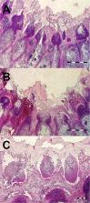
          Although laboratory rabbits are commonly used as models of appendicitis in man, spontaneous appendicitis was only described ante-mortem in one pet rabbit with an acute abdomen. The aim of this article is to describe two spontaneous cases of appendicitis in pet rabbits, to describe therapeutic appendectomy, and to discuss the microbial flora of the inflamed appendix. A 5-month-old intact female and a 16-month-old, neutered male were presented to the veterinary clinic with restlessness, anorexia, and reduced faecal output. The main clinical findings were restlessness, severe discomfort on abdominal palpation, a mid-abdominal palpable tubulous mass and an elevated rectal temperature. Blood analyses showed lymphocytosis, monocytosis, and hyperglycaemia. Radiography was inconclusive. Abdominal ultrasound revealed a presence of a tubular structure with wall thicknesses of 4.2 and 3.7 mm in the two rabbits, respectively. The tubular structure had a rounded, closed end, and a multilayered wall, suggestive of appendicitis. Due to metabolic acidosis and poor prognosis, the first rabbit was euthanized. In the 16-month-old rabbit, appendectomy was performed. Recovery was uneventful, and 4 h after surgery, the rabbit started to become normally active. Postoperative care consisted of fluid therapy, multimodal analgesia, supportive care and prokinetics. Follow-up examinations at 10 days, 1 month, and at 11 months after the surgery did not show any abnormal clinical or laboratory findings. Histopathological examination of appendices from both rabbits showed gangrenous appendicitis. Aerobic cultivation showed the presence of pure culture of Klebsiella variicola sensitive to enrofloxacin, marbofloxacin, tetracycline, cefuroxime, trimethoprim sulphonamide, neomycin, and gentamicin. Restlessness associated with anorexia, abdominal pain, palpable abdominal mass, hyperglycaemia, lymphocytosis, and elevated rectal temperature may be indicative of inflammation within the gastrointestinal tract. Abdominal ultrasound is recommended in rabbits with showing these clinical signs because radiography can be inconclusive. Appendicitis is a life-threatening condition, which should be included into the list of differential diagnoses; for the rabbit, an acute abdomen and gastrointestinal stasis syndrome and must be treated immediately. K. variicola may be associated with appendicitis in rabbits as a causative agent or in association with appendix intraluminal dysmicrobia.

          Related collections

          Most cited references35

          • Record: found
          • Abstract: found
          • Article: not found

          Acute appendicitis: modern understanding of pathogenesis, diagnosis, and management.

          Acute appendicitis is one of the most common abdominal emergencies worldwide. The cause remains poorly understood, with few advances in the past few decades. To obtain a confident preoperative diagnosis is still a challenge, since the possibility of appendicitis must be entertained in any patient presenting with an acute abdomen. Although biomarkers and imaging are valuable adjuncts to history and examination, their limitations mean that clinical assessment is still the mainstay of diagnosis. A clinical classification is used to stratify management based on simple (non-perforated) and complex (gangrenous or perforated) inflammation, although many patients remain with an equivocal diagnosis, which is one of the most challenging dilemmas. An observed divide in disease course suggests that some cases of simple appendicitis might be self-limiting or respond to antibiotics alone, whereas another type often seems to perforate before the patient reaches hospital. Although the mortality rate is low, postoperative complications are common in complex disease. We discuss existing knowledge in pathogenesis, modern diagnosis, and evolving strategies in management that are leading to stratified care for patients.
            Bookmark
            • Record: found
            • Abstract: found
            • Article: found
            Is Open Access

            Identification of Klebsiella pneumoniae, Klebsiella quasipneumoniae, Klebsiella variicola and Related Phylogroups by MALDI-TOF Mass Spectrometry

            Klebsiella pneumoniae (phylogroup Kp1), one of the most problematic pathogens associated with antibiotic resistance worldwide, is phylogenetically closely related to K. quasipneumoniae [subsp. quasipneumoniae (Kp2) and subsp. similipneumoniae (Kp4)], K. variicola (Kp3) and two unnamed phylogroups (Kp5 and Kp6). Together, Kp1 to Kp6 make-up the K. pneumoniae complex. Currently, the phylogroups can be reliably identified only based on gene (or genome) sequencing. Misidentification using standard laboratory methods is common and consequently, the clinical significance of K. pneumoniae complex members is imprecisely defined. Here, we evaluated and validated the potential of MALDI-TOF mass spectrometry (MS) to discriminate K. pneumoniae complex members. We detected mass spectrometry biomarkers associated with the phylogroups, with a sensitivity and specificity ranging between 80–100% and 97–100%, respectively. Strains within phylogroups Kp1, Kp2, Kp4, and Kp5 each shared two specific peaks not observed in other phylogroups. Kp3 strains shared a peak that was only observed otherwise in Kp5. Finally, Kp6 had a diagnostic peak shared only with Kp1. Kp3 and Kp6 could therefore be identified by exclusion criteria (lacking Kp5 and Kp1-specific peaks, respectively). Further, ranked Pearson correlation clustering of spectra grouped strains according to their phylogroup. The model was tested and successfully validated using different culture media. These results demonstrate the potential of MALDI-TOF MS for precise identification of K. pneumoniae complex members. Incorporation of spectra of all K. pneumoniae complex members into reference MALDI-TOF spectra databases, in which they are currently lacking, is desirable. MALDI-TOF MS may thereby enable a better understanding of the epidemiology, ecology, and pathogenesis of members of the K. pneumoniae complex.
              Bookmark
              • Record: found
              • Abstract: found
              • Article: found
              Is Open Access

              Antimicrobial use in food animals and human health: time to implement ‘One Health’ approach

              The use of antimicrobials in animals for growth promotion and infection prevention significantly contributes to the development of antimicrobial resistance (AMR), a growing public health threat. While the World Health Organization (WHO), the United Nations (UN) and the European Union (EU) have taken steps towards reducing and restricting the use of antimicrobials in animals, initiatives are insufficient in developing countries where the demands for food animals continue to rise over the years. The inter-sectoral acknowledgment of inextricable link between animal health, human health and the environment (One Health approach) is critical. Concerted and collaborative efforts among all the stakeholders are essential to deal with this complex problem of resistance.
                Bookmark

                Author and article information

                Contributors
                Journal
                Front Vet Sci
                Front Vet Sci
                Front. Vet. Sci.
                Frontiers in Veterinary Science
                Frontiers Media S.A.
                2297-1769
                08 December 2021
                2021
                : 8
                : 779517
                Affiliations
                [1] 1Department of Pharmacology and Pharmacy, Faculty of Veterinary Medicine, University of Veterinary Sciences Brno , Brno, Czechia
                [2] 2Jekl & Hauptman Veterinary Clinic , Brno, Czechia
                [3] 3Department of Pathological Morphology, Faculty of Veterinary Medicine, Veterinary University , Brno, Czechia
                Author notes

                Edited by: Tanmoy Rana, West Bengal University of Animal and Fishery Sciences, India

                Reviewed by: Dharm Singh Meena, Post-Graduate Institute of Veterinary Education and Research (PGIVER), India; Saravanan Mani, Tamil Nadu Veterinary and Animal Sciences University, India

                *Correspondence: Vladimir Jekl vladimirjekl@ 123456gmail.com

                This article was submitted to Veterinary Emergency and Critical Care Medicine, a section of the journal Frontiers in Veterinary Science

                Article
                10.3389/fvets.2021.779517
                8692788
                ece5504e-5979-4664-a9d0-49ea144590e5
                Copyright © 2021 Jekl, Piskovska, Drnkova, Skoric, Hauptman and Chloupek.

                This is an open-access article distributed under the terms of the Creative Commons Attribution License (CC BY). The use, distribution or reproduction in other forums is permitted, provided the original author(s) and the copyright owner(s) are credited and that the original publication in this journal is cited, in accordance with accepted academic practice. No use, distribution or reproduction is permitted which does not comply with these terms.

                History
                : 18 September 2021
                : 08 November 2021
                Page count
                Figures: 3, Tables: 1, Equations: 0, References: 37, Pages: 7, Words: 5235
                Funding
                Funded by: Veterinární a Farmaceutická Univerzita Brno, doi 10.13039/501100004755;
                Categories
                Veterinary Science
                Case Report

                rabbit,appendix,appendicitis,gastrointestinal stasis,acute abdomen,klebsiella

                Comments

                Comment on this article