33
views
0
recommends
+1 Recommend
0 collections
    0
    shares
      • Record: found
      • Abstract: found
      • Article: found
      Is Open Access

      EGFR, CD10 and proliferation marker Ki67 expression in ameloblastoma: possible role in local recurrence

      research-article
      1 , , 1
      Diagnostic Pathology
      BioMed Central
      Ameloblastoma, EGFR, CD10, Ki67 and recurrence

      Read this article at

      Bookmark
          There is no author summary for this article yet. Authors can add summaries to their articles on ScienceOpen to make them more accessible to a non-specialist audience.

          Abstract

          Background

          Ameloblastoma is an odontogenic neoplasm characterized by local invasiveness and tendency towards recurrence.

          Aims

          Studying the role played by EGFR, CD10 and Ki67 in the recurrence of ameloblastoma.

          Methods

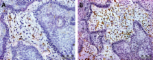
          This study was carried out on 22 retrospective cases of mandibular ameloblastoma from the period from Jan 2002 to Jan 2008 with follow up period until Jan 2011 (3 to 8 years follow up peroid). Archival materials were obtained from pathology department, Mansoura university. Paraffin sections of tumor tissue from all cases were submitted for routine H&E stains and immunohistochemistry using EGFR, CD10 and Ki67 monoclonal antibodies. Statistical analysis using of clinical data for all patients, tumor type, EGFR, CD10 and Ki67 expression in relation to recurrence were evaluated.

          Results

          Among the 22 cases, 10 cases were males and 12 were females with sex ratio 1:1.2. Age ranged from 34 to 59 years old with a mean age 44.18 year. Five cases showed local recurrence within studied period and proved by biopsy. No statistically significant relation was found between local recurrence and patient age, tumor size, tumor type, EGFR expression. There was a significant relation between CD10 expression as well as Ki67 labelling index and recurrence (P value = 0.003, 0.000 respectively).

          Conclusion

          Evaluation of CD10 and Ki67 status together with conventional histological evaluation can help in providing more information about the biologic behavior of the tumor, while EGFR could be a target of an expanding class of anticancer therapies.

          Since ameloblastomas are EGFR-positive tumors, anti-EGFR agents could be considered to reduce the size of large tumors and to treat unresectable tumors that are in close proximity to vital structures.

          Virtual Slides

          The virtual slide(s) for this article can be found here:

          http://www.diagnosticpathology.diagnomx.eu/vs/1902106905645651

          Related collections

          Most cited references33

          • Record: found
          • Abstract: found
          • Article: not found

          A comprehensive pathway map of epidermal growth factor receptor signaling

          The epidermal growth factor receptor (EGFR) signaling pathway is one of the most important pathways that regulate growth, survival, proliferation, and differentiation in mammalian cells. Reflecting this importance, it is one of the best-investigated signaling systems, both experimentally and computationally, and several computational models have been developed for dynamic analysis. A map of molecular interactions of the EGFR signaling system is a valuable resource for research in this area. In this paper, we present a comprehensive pathway map of EGFR signaling and other related pathways. The map reveals that the overall architecture of the pathway is a bow-tie (or hourglass) structure with several feedback loops. The map is created using CellDesigner software that enables us to graphically represent interactions using a well-defined and consistent graphical notation, and to store it in Systems Biology Markup Language (SBML).
            Bookmark
            • Record: found
            • Abstract: not found
            • Article: not found

            EGF mutant receptor vIII as a molecular target in cancer therapy.

              Bookmark
              • Record: found
              • Abstract: found
              • Article: not found

              Odontogenic cysts, odontogenic tumors, fibroosseous, and giant cell lesions of the jaws.

              J Regezi (2002)
              Odontogenic cysts that can be problematic because of recurrence and/or aggressive growth include odontogenic keratocyst (OKC), calcifying odontogenic cyst, and the recently described glandular odontogenic cyst. The OKC has significant growth capacity and recurrence potential and is occasionally indicative of the nevoid basal cell carcinoma syndrome. There is also an orthokeratinized variant, the orthokeratinized odontogenic cyst, which is less aggressive and is not syndrome associated. Ghost cell keratinization, which typifies the calcifying odontogenic cyst, can be seen in solid lesions that have now been designated odontogenic ghost cell tumor. The glandular odontogenic cyst contains mucous cells and ductlike structures that may mimic central mucoepidermoid carcinoma. Several odontogenic tumors may provide diagnostic challenges, particularly the cystic ameloblastoma. Identification of this frequently underdiagnosed cystic tumor often comes after one or more recurrences and a destructive course. Other difficult lesions include malignant ameloblastomas, calcifying epithelial odontogenic tumor, squamous odontogenic tumor, and clear-cell odontogenic tumor. Histologic identification of myxofibrous lesions of the jaws (odontogenic myxoma, odontogenic fibroma, desmoplastic fibroma) is necessary to avoid the diagnostic pitfall of overdiagnosis of similar-appearing follicular sacs and dental pulps. Fibroosseous lesions of the jaws show considerable microscopic overlap and include fibrous dysplasia, ossifying fibroma, periapical cementoosseous dysplasia, and low-grade chronic osteomyelitis. The term fibrous dysplasia is probably overused in general practice and should be reserved for the rare lesion that presents as a large, expansile, diffuse opacity of children and young adults. The need to use clinicopathologic correlation in assessing these lesions is of particular importance. Central giant cell granuloma is a relatively common jaw lesion of young adults that has an unpredictable behavior. Microscopic diagnosis is relatively straightforward; however, this lesion continues to be somewhat controversial because of its disputed classification (reactive versus neoplastic) and because of its management (surgical versus. medical). Its relationship to giant cell tumor of long bone remains undetermined.
                Bookmark

                Author and article information

                Journal
                Diagn Pathol
                Diagn Pathol
                Diagnostic Pathology
                BioMed Central
                1746-1596
                2012
                2 February 2012
                : 7
                : 14
                Affiliations
                [1 ]Pathology Department, Faculty of medicine, Mansoura University, Egypt
                Article
                1746-1596-7-14
                10.1186/1746-1596-7-14
                3328247
                22300665
                ed17b33d-c108-4916-85f7-4d91dfea4d96
                Copyright ©2012 Abdel-Aziz and Amin; licensee BioMed Central Ltd.

                This is an Open Access article distributed under the terms of the Creative Commons Attribution License ( http://creativecommons.org/licenses/by/2.0), which permits unrestricted use, distribution, and reproduction in any medium, provided the original work is properly cited.

                History
                : 5 December 2011
                : 2 February 2012
                Categories
                Research

                Pathology
                ameloblastoma,ki67 and recurrence,cd10,egfr
                Pathology
                ameloblastoma, ki67 and recurrence, cd10, egfr

                Comments

                Comment on this article