23
views
0
recommends
+1 Recommend
0 collections
    0
    shares
      • Record: found
      • Abstract: found
      • Article: found
      Is Open Access

      Sonographically Guided Core Biopsy of the Breast: Comparison of 14-Gauge Automated Gun and 11-Gauge Directional Vacuum-Assisted Biopsy Methods

      research-article

      Read this article at

      Bookmark
          There is no author summary for this article yet. Authors can add summaries to their articles on ScienceOpen to make them more accessible to a non-specialist audience.

          Abstract

          Objective

          To compare the outcomes of 14-gauge automated biopsy and 11-gauge vacuum-assisted biopsy for the sonographically guided core biopsies of breast lesions.

          Materials and Methods

          We retrospectively reviewed all sonographically guided core biopsies performed from January 2002 to February 2004. The sonographically guided core biopsies were performed with using a 14-gauge automated gun on 562 breast lesions or with using an 11-gauge vacuum-assisted device on 417 lesions. The histologic findings were compared with the surgical, imaging and follow-up findings. The histologic underestimation rate, the repeat biopsy rate and the false negative rates were compared between the two groups.

          Results

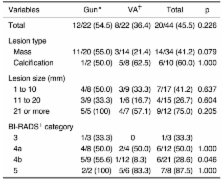
          A repeat biopsy was performed on 49 benign lesions because of the core biopsy results of the high-risk lesions (n = 24), the imaging-histologic discordance (n = 5), and the imaging findings showing disease progression (n = 20). The total underestimation rates, according to the biopsy device, were 55% (12/22) for the 14-gauge automated gun biopsies and 36% (8/22) for the 11-gauge vacuum-assisted device ( p = 0.226). The atypical ductal hyperplasia (ADH) underestimation (i.e., atypical ductal hyperplasia at core biopsy and carcinoma at surgery) was 58% (7/12) for the 14-gauge automated gun biopsies and 20% (1/5) for the 11-gauge vacuum-assisted biopsies. The ductal carcinoma in situ (DCIS) underestimation rate (i.e., ductal carcinoma in situ upon core biopsy and invasive carcinoma found at surgery) was 50% (5/10) for the 14-gauge automated gun biopsies and 41% (7/17) for the 11-gauge vacuum-assisted biopsies. The repeat biopsy rates were 6% (33/562) for the 14-gauge automated gun biopsies and 3.5% (16/417) for the 11-gauge vacuum-assisted biopsies. Only 5 (0.5%) of the 979 core biopsies were believed to have missed the malignant lesions. The false-negative rate was 3% (4 of 128 cancers) for the 14-gauge automated gun biopsies and 1% (1 of 69 cancers) for the 11-gauge vacuum-assisted biopsies.

          Conclusion

          The outcomes of the sonographically guided core biopsies performed with the 11-gauge vacuum-assisted device were better than those outcomes of the biopsies performed with the 14-gauge automated gun in terms of underestimation, rebiopsy and the false negative rate, although these differences were not statistically significant.

          Related collections

          Most cited references29

          • Record: found
          • Abstract: found
          • Article: not found

          Percutaneous large-core breast biopsy: a multi-institutional study.

          To assess the reliability and reproducibility of automated large-core breast biopsy. A consortium of 20 institutions reported, in a standardized fashion, their core breast biopsy data. All biopsies were performed with "long-throw" (2.3-cm) automated core biopsy devices fitted with 14-gauge needles. Needle guidance was accomplished by means of either a dedicated, stereotaxic device, in which the patient lies in the prone position, or high-frequency electronically focused ultrasound equipment. The data in 6,152 lesions were gathered. Clinical or surgical follow-up was available in 3,765 lesions; 1,363 of these lesions were subsequently surgically excised, and the core histologic study showed cancer in 910 lesions, mammary intraepithelial neoplasia in 173 lesions, and benign disease in 280 lesions. In these 280 lesions, there were 15 false-negative core biopsies. The data show that percutaneous large-core breast biopsy is a reproducible and reliable alternative to surgical biopsy.
            Bookmark
            • Record: found
            • Abstract: found
            • Article: not found

            US-guided automated large-core breast biopsy.

            To evaluate the efficacy of ultrasound (US)-guided automated large-core percutaneous needle breast biopsy, biopsy of 181 ultrasonographically suspicious breast lesions was performed by using a long-throw biopsy gun and 14-gauge needles with continuous US guidance. A "freehand" technique with either 5.0- or 7.5-MHz linear-array transducers was used. Needle core diagnoses were compared with surgical diagnoses in the 49 lesions subsequently surgically excised. The remaining 132 cases were followed for 12-36 months. Agreement between needle-core and surgical diagnoses in the 49 lesions was 100%. This group included 34 cancers (28 infiltrating ductal, two mucinous, one mixed infiltrating ductal and lobular, and one each of intraductal, infiltrating lobular, and tubular carcinoma). To date, no cancers have been found in the other 132 cases. The procedure time averaged 20 minutes, and no complications occurred. With an accuracy approaching that of excisional biopsy, US-guided needle core biopsy provides an alternative to surgery.
              Bookmark
              • Record: found
              • Abstract: found
              • Article: not found

              Stereotactic breast biopsy of nonpalpable lesions: determinants of ductal carcinoma in situ underestimation rates.

              To measure the effect of biopsy device, probe size, mammographic lesion type, lesion size, and number of samples obtained per lesion on the ductal carcinoma in situ (DCIS) underestimation rate. Nonpalpable breast lesions at 16 institutions received a histologic diagnosis of DCIS after 14-gauge automated large-core biopsy in 373 lesions and after 14- or 11-gauge directional vacuum-assisted biopsy in 953 lesions. The presence of histopathologic invasive carcinoma was noted at subsequent surgical biopsy. By performing the chi(2) test, independent significant DCIS underestimation rates by biopsy device were 20.4% (76 of 373) of lesions diagnosed at large-core biopsy and 11.2% (107 of 953) of lesions diagnosed at vacuum-assisted biopsy (P <.001); by lesion type, 24.3% (35 of 144) of masses and 12.5% (148 of 1,182) of microcalcifications (P <.001); and by number of specimens per lesion, 17.5% (88 of 502) with 10 or fewer specimens and 11.5% (92 of 799) with greater than 10 (P <.02). DCIS underestimations increased with lesion size. DCIS underestimations were 1.9 times more frequent with masses than with calcifications, 1.8 times more frequent with large-core biopsy than with vacuum-assisted biopsy, and 1.5 times more frequent with 10 or fewer specimens per lesion than with more than 10 specimens per lesion.
                Bookmark

                Author and article information

                Journal
                Korean J Radiol
                KJR
                Korean Journal of Radiology
                The Korean Radiological Society
                1229-6929
                2005-8330
                Apr-Jun 2005
                30 June 2005
                : 6
                : 2
                : 102-109
                Affiliations
                [1 ]Department of Radiology, Seoul National University College of Medicine, The Institute of Radiation Medicine, SNUMRC, and The Clinical Research Institute, Seoul National University Hospital, Korea.
                [2 ]Department of Pathology, Seoul National University College of Medicine, Korea.
                [3 ]Department of Surgery, Seoul National University College of Medicine, Korea.
                Author notes
                Address reprint requests to: Woo Kyung Moon, MD, Department of Diagnostic Radiology, Seoul National University Hospital, 28 Yongon-dong, Chongno-gu, Seoul 110-744, Korea. Tel. (822) 2072-3928, Fax. (822) 747-7418, moonwk@ 123456radcom.snu.ac.kr
                Article
                10.3348/kjr.2005.6.2.102
                2686400
                15968149
                ed524cfb-7729-41df-8c5f-d50c22179c95
                Copyright © 2005 The Korean Radiological Society

                This is an Open Access article distributed under the terms of the Creative Commons Attribution Non-Commercial License ( http://creativecommons.org/licenses/by-nc/3.0) which permits unrestricted non-commercial use, distribution, and reproduction in any medium, provided the original work is properly cited.

                History
                : 19 October 2004
                : 09 February 2005
                Categories
                Original Article

                Radiology & Imaging
                breast, us,breast, biopsy,biopsies, technology,breast neoplasms, diagnosis
                Radiology & Imaging
                breast, us, breast, biopsy, biopsies, technology, breast neoplasms, diagnosis

                Comments

                Comment on this article