2
views
0
recommends
+1 Recommend
0 collections
    0
    shares
      • Record: found
      • Abstract: found
      • Article: found
      Is Open Access

      Patient-specific modeling for guided rehabilitation of stroke patients: the BrainX3 use-case

      brief-report

      Read this article at

      Bookmark
          There is no author summary for this article yet. Authors can add summaries to their articles on ScienceOpen to make them more accessible to a non-specialist audience.

          Abstract

          BrainX3 is an interactive neuroinformatics platform that has been thoughtfully designed to support neuroscientists and clinicians with the visualization, analysis, and simulation of human neuroimaging, electrophysiological data, and brain models. The platform is intended to facilitate research and clinical use cases, with a focus on personalized medicine diagnostics, prognostics, and intervention decisions. BrainX3 is designed to provide an intuitive user experience and is equipped to handle different data types and 3D visualizations. To enhance patient-based analysis, and in keeping with the principles of personalized medicine, we propose a framework that can assist clinicians in identifying lesions and making patient-specific intervention decisions. To this end, we are developing an AI-based model for lesion identification, along with a mapping of tract information. By leveraging the patient's lesion information, we can gain valuable insights into the structural damage caused by the lesion. Furthermore, constraining whole-brain models with patient-specific disconnection masks can allow for the detection of mesoscale excitatory-inhibitory imbalances that cause disruptions in macroscale network properties. Finally, such information has the potential to guide neuromodulation approaches, assisting in the choice of candidate targets for stimulation techniques such as Transcranial Ultrasound Stimulation (TUS), which modulate E-I balance, potentiating cortical reorganization and the restoration of the dynamics and functionality disrupted due to the lesion.

          Related collections

          Most cited references106

          • Record: found
          • Abstract: not found
          • Book Chapter: not found

          U-Net: Convolutional Networks for Biomedical Image Segmentation

            Bookmark
            • Record: found
            • Abstract: found
            • Article: found
            Is Open Access

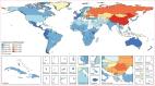
            Global, regional, and national burden of stroke, 1990–2016: a systematic analysis for the Global Burden of Disease Study 2016

            Summary Background Stroke is a leading cause of mortality and disability worldwide and the economic costs of treatment and post-stroke care are substantial. The Global Burden of Diseases, Injuries, and Risk Factors Study (GBD) provides a systematic, comparable method of quantifying health loss by disease, age, sex, year, and location to provide information to health systems and policy makers on more than 300 causes of disease and injury, including stroke. The results presented here are the estimates of burden due to overall stroke and ischaemic and haemorrhagic stroke from GBD 2016. Methods We report estimates and corresponding uncertainty intervals (UIs), from 1990 to 2016, for incidence, prevalence, deaths, years of life lost (YLLs), years lived with disability (YLDs), and disability-adjusted life-years (DALYs). DALYs were generated by summing YLLs and YLDs. Cause-specific mortality was estimated using an ensemble modelling process with vital registration and verbal autopsy data as inputs. Non-fatal estimates were generated using Bayesian meta-regression incorporating data from registries, scientific literature, administrative records, and surveys. The Socio-demographic Index (SDI), a summary indicator generated using educational attainment, lagged distributed income, and total fertility rate, was used to group countries into quintiles. Findings In 2016, there were 5·5 million (95% UI 5·3 to 5·7) deaths and 116·4 million (111·4 to 121·4) DALYs due to stroke. The global age-standardised mortality rate decreased by 36·2% (−39·3 to −33·6) from 1990 to 2016, with decreases in all SDI quintiles. Over the same period, the global age-standardised DALY rate declined by 34·2% (−37·2 to −31·5), also with decreases in all SDI quintiles. There were 13·7 million (12·7 to 14·7) new stroke cases in 2016. Global age-standardised incidence declined by 8·1% (−10·7 to −5·5) from 1990 to 2016 and decreased in all SDI quintiles except the middle SDI group. There were 80·1 million (74·1 to 86·3) prevalent cases of stroke globally in 2016; 41·1 million (38·0 to 44·3) in women and 39·0 million (36·1 to 42·1) in men. Interpretation Although age-standardised mortality rates have decreased sharply from 1990 to 2016, the decrease in age-standardised incidence has been less steep, indicating that the burden of stroke is likely to remain high. Planned updates to future GBD iterations include generating separate estimates for subarachnoid haemorrhage and intracerebral haemorrhage, generating estimates of transient ischaemic attack, and including atrial fibrillation as a risk factor. Funding Bill & Melinda Gates Foundation
              Bookmark
              • Record: found
              • Abstract: found
              • Article: not found

              Efficient multi-scale 3D CNN with fully connected CRF for accurate brain lesion segmentation

              We propose a dual pathway, 11-layers deep, three-dimensional Convolutional Neural Network for the challenging task of brain lesion segmentation. The devised architecture is the result of an in-depth analysis of the limitations of current networks proposed for similar applications. To overcome the computational burden of processing 3D medical scans, we have devised an efficient and effective dense training scheme which joins the processing of adjacent image patches into one pass through the network while automatically adapting to the inherent class imbalance present in the data. Further, we analyze the development of deeper, thus more discriminative 3D CNNs. In order to incorporate both local and larger contextual information, we employ a dual pathway architecture that processes the input images at multiple scales simultaneously. For post-processing of the network's soft segmentation, we use a 3D fully connected Conditional Random Field which effectively removes false positives. Our pipeline is extensively evaluated on three challenging tasks of lesion segmentation in multi-channel MRI patient data with traumatic brain injuries, brain tumours, and ischemic stroke. We improve on the state-of-the-art for all three applications, with top ranking performance on the public benchmarks BRATS 2015 and ISLES 2015. Our method is computationally efficient, which allows its adoption in a variety of research and clinical settings. The source code of our implementation is made publicly available.
                Bookmark

                Author and article information

                Contributors
                URI : http://loop.frontiersin.org/people/2412903/overviewRole:
                URI : http://loop.frontiersin.org/people/1501205/overviewRole: Role:
                URI : http://loop.frontiersin.org/people/5803/overviewRole: Role:
                Journal
                Front Neurol
                Front Neurol
                Front. Neurol.
                Frontiers in Neurology
                Frontiers Media S.A.
                1664-2295
                30 November 2023
                2023
                : 14
                : 1279875
                Affiliations
                [1] 1Donders Institute for Brain, Cognition and Behavior, Radboud University , Nijmegen, Netherlands
                [2] 2Eodyne Systems S.L. , Barcelona, Spain
                [3] 3Department of Information and Communication Technologies, Universitat Pompeu Fabra (UPF) , Barcelona, Spain
                Author notes

                Edited by: Spase Petkoski, Aix Marseille Université, France

                Reviewed by: Edgar Hernandez, National University of Colombia, Colombia

                *Correspondence: Vivek Sharma vivek.sharma@ 123456donders.ru.nl

                †These authors share first authorship

                Article
                10.3389/fneur.2023.1279875
                10719856
                38099071
                edfe1f1d-59f2-41b0-b69d-e3ac0dce5110
                Copyright © 2023 Sharma, Páscoa dos Santos and Verschure.

                This is an open-access article distributed under the terms of the Creative Commons Attribution License (CC BY). The use, distribution or reproduction in other forums is permitted, provided the original author(s) and the copyright owner(s) are credited and that the original publication in this journal is cited, in accordance with accepted academic practice. No use, distribution or reproduction is permitted which does not comply with these terms.

                History
                : 22 August 2023
                : 06 November 2023
                Page count
                Figures: 2, Tables: 0, Equations: 0, References: 108, Pages: 9, Words: 7057
                Funding
                The author(s) declare financial support was received for the research, authorship, and/or publication of this article. This work was funded by the European Commission's Counterfactual Assessment and Valuation for Awareness Architecture-CAVAA (European Commission, EIC 101071178), AI in Stroke Neurorehabilitation-AISN (European Commission, EIC 101057655), Personalised Health cognitive assistance for RehAbilitation SystEm-PHRASE (European Commission, EIC 101058240), eBRAIN-Health (European Commission, EIC 101058516) and European School of Network Neuroscience-euSNN (MSCA-ITN ETN H2020-ID 860563). The funders had no role in the conceptualization, analysis, decision to publish, or preparation of the manuscript.
                Categories
                Neurology
                Perspective
                Custom metadata
                Neurorehabilitation

                Neurology
                automatic lesion identification,whole-brain models,transcranial ultrasound stimulation,brainx3,stroke

                Comments

                Comment on this article