32
views
0
recommends
+1 Recommend
0 collections
    0
    shares
      • Record: found
      • Abstract: found
      • Article: found
      Is Open Access

      Dual-color STED microscopy reveals a sandwich structure of Bassoon and Piccolo in active zones of adult and aged mice

      research-article

      Read this article at

      Bookmark
          There is no author summary for this article yet. Authors can add summaries to their articles on ScienceOpen to make them more accessible to a non-specialist audience.

          Abstract

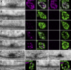
          Presynaptic active zones play a pivotal role as synaptic vesicle release sites for synaptic transmission, but the molecular architecture of active zones in mammalian neuromuscular junctions (NMJs) at sub-diffraction limited resolution remains unknown. Bassoon and Piccolo are active zone specific cytosolic proteins essential for active zone assembly in NMJs, ribbon synapses, and brain synapses. These proteins are thought to colocalize and share some functions at active zones. Here, we report an unexpected finding of non-overlapping localization of these two proteins in mouse NMJs revealed using dual-color stimulated emission depletion (STED) super resolution microscopy. Piccolo puncta sandwiched Bassoon puncta and aligned in a Piccolo-Bassoon-Piccolo structure in adult NMJs. P/Q-type voltage-gated calcium channel (VGCC) puncta colocalized with Bassoon puncta. The P/Q-type VGCC and Bassoon protein levels decreased significantly in NMJs from aged mouse. In contrast, the Piccolo levels in NMJs from aged mice were comparable to levels in adult mice. This study revealed the molecular architecture of active zones in mouse NMJs at sub-diffraction limited resolution, and described the selective degeneration mechanism of active zone proteins in NMJs from aged mice. Interestingly, the localization pattern of active zone proteins described herein is similar to active zone structures described using electron microscope tomography.

          Related collections

          Most cited references55

          • Record: found
          • Abstract: found
          • Article: not found

          Superresolution imaging of chemical synapses in the brain.

          Determination of the molecular architecture of synapses requires nanoscopic image resolution and specific molecular recognition, a task that has so far defied many conventional imaging approaches. Here, we present a superresolution fluorescence imaging method to visualize the molecular architecture of synapses in the brain. Using multicolor, three-dimensional stochastic optical reconstruction microscopy, the distributions of synaptic proteins can be measured with nanometer precision. Furthermore, the wide-field, volumetric imaging method enables high-throughput, quantitative analysis of a large number of synapses from different brain regions. To demonstrate the capabilities of this approach, we have determined the organization of ten protein components of the presynaptic active zone and the postsynaptic density. Variations in synapse morphology, neurotransmitter receptor composition, and receptor distribution were observed both among synapses and across different brain regions. Combination with optogenetics further allowed molecular events associated with synaptic plasticity to be resolved at the single-synapse level. Copyright © 2010 Elsevier Inc. All rights reserved.
            Bookmark
            • Record: found
            • Abstract: found
            • Article: found
            Is Open Access

            Maturation of active zone assembly by Drosophila Bruchpilot

            Synaptic vesicles fuse at active zone (AZ) membranes where Ca2+ channels are clustered and that are typically decorated by electron-dense projections. Recently, mutants of the Drosophila melanogaster ERC/CAST family protein Bruchpilot (BRP) were shown to lack dense projections (T-bars) and to suffer from Ca2+ channel–clustering defects. In this study, we used high resolution light microscopy, electron microscopy, and intravital imaging to analyze the function of BRP in AZ assembly. Consistent with truncated BRP variants forming shortened T-bars, we identify BRP as a direct T-bar component at the AZ center with its N terminus closer to the AZ membrane than its C terminus. In contrast, Drosophila Liprin-α, another AZ-organizing protein, precedes BRP during the assembly of newly forming AZs by several hours and surrounds the AZ center in few discrete punctae. BRP seems responsible for effectively clustering Ca2+ channels beneath the T-bar density late in a protracted AZ formation process, potentially through a direct molecular interaction with intracellular Ca2+ channel domains.
              Bookmark
              • Record: found
              • Abstract: found
              • Article: not found

              The presynaptic active zone protein bassoon is essential for photoreceptor ribbon synapse formation in the retina.

              The photoreceptor ribbon synapse is a highly specialized glutamatergic synapse designed for the continuous flow of synaptic vesicles to the neurotransmitter release site. The molecular mechanisms underlying ribbon synapse formation are poorly understood. We have investigated the role of the presynaptic cytomatrix protein Bassoon, a major component of the photoreceptor ribbon, in a mouse retina deficient of functional Bassoon protein. Photoreceptor ribbons lacking Bassoon are not anchored to the presynaptic active zones. This results in an impaired photoreceptor synaptic transmission, an abnormal dendritic branching of neurons postsynaptic to photoreceptors, and the formation of ectopic synapses. These findings suggest a critical role of Bassoon in the formation and the function of photoreceptor ribbon synapses of the mammalian retina.
                Bookmark

                Author and article information

                Journal
                Sci Rep
                Sci Rep
                Scientific Reports
                Nature Publishing Group
                2045-2322
                20 June 2016
                2016
                : 6
                : 27935
                Affiliations
                [1 ]Department of Anatomy and Cell Biology, University of Kansas School of Medicine , Kansas City, 66160, USA
                [2 ]Department of Geriatric Medicine, Tokyo Metropolitan Institute of Gerontology , Itabashi-ku, Tokyo 173-0015, Japan
                Author notes
                [*]

                These authors contributed equally to this work.

                Article
                srep27935
                10.1038/srep27935
                4913281
                27321892
                ee48c57f-2cfd-49bb-b130-f5229abc2b72
                Copyright © 2016, Macmillan Publishers Limited

                This work is licensed under a Creative Commons Attribution 4.0 International License. The images or other third party material in this article are included in the article’s Creative Commons license, unless indicated otherwise in the credit line; if the material is not included under the Creative Commons license, users will need to obtain permission from the license holder to reproduce the material. To view a copy of this license, visit http://creativecommons.org/licenses/by/4.0/

                History
                : 04 February 2016
                : 16 May 2016
                Categories
                Article

                Uncategorized
                Uncategorized

                Comments

                Comment on this article