1
views
0
recommends
+1 Recommend
0 collections
    0
    shares
      • Record: found
      • Abstract: found
      • Article: found
      Is Open Access

      IL-17: an important pathogenic factor in endometriosis

      review-article

      Read this article at

      Bookmark
          There is no author summary for this article yet. Authors can add summaries to their articles on ScienceOpen to make them more accessible to a non-specialist audience.

          Abstract

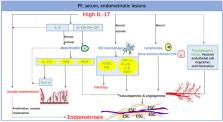
          Interleukin-17 (IL-17) is known as a Th17-cell-derived proinflammatory cytokine, which plays a pivotal role in several inflammatory and autoimmune diseases such as systemic lupus erythematosus (SLE), rheumatoid arthritis, and psoriasis. Emerging evidence has shown that IL-17 is linked to endometriosis, although the etiology of endometriosis is still unknown. The IL-17 expression is up-regulated in serum, peritoneal fluid (PF) and endometriotic lesions from patients with endometriosis but the related regulation mechanisms are complex and obscure. Meanwhile, the specific roles of IL-17 in endometriosis are also worthy of further exploration. Through the integration and summary of literature, we conclude that the secretion of IL-17 increases under the regulation of ectopic microenvironment and other factors, and then IL-17 is deeply involved in endometriosis in the regulation of immune microenvironment, the invasion and growth of ectopic lesions, and so on, which implies its therapeutic value in this disorder.

          Related collections

          Most cited references145

          • Record: found
          • Abstract: found
          • Article: not found

          A distinct lineage of CD4 T cells regulates tissue inflammation by producing interleukin 17.

          Interleukin 17 (IL-17) has been linked to autoimmune diseases, although its regulation and function have remained unclear. Here we have evaluated in vitro and in vivo the requirements for the differentiation of naive CD4 T cells into effector T helper cells that produce IL-17. This process required the costimulatory molecules CD28 and ICOS but was independent of the cytokines and transcription factors required for T helper type 1 or type 2 differentiation. Furthermore, both IL-4 and interferon-gamma negatively regulated T helper cell production of IL-17 in the effector phase. In vivo, antibody to IL-17 inhibited chemokine expression in the brain during experimental autoimmune encephalomyelitis, whereas overexpression of IL-17 in lung epithelium caused chemokine production and leukocyte infiltration. Thus, IL-17 expression characterizes a unique T helper lineage that regulates tissue inflammation.
            Bookmark
            • Record: found
            • Abstract: found
            • Article: not found

            Macrophages in Tissue Repair, Regeneration, and Fibrosis.

            Inflammatory monocytes and tissue-resident macrophages are key regulators of tissue repair, regeneration, and fibrosis. After tissue injury, monocytes and macrophages undergo marked phenotypic and functional changes to play critical roles during the initiation, maintenance, and resolution phases of tissue repair. Disturbances in macrophage function can lead to aberrant repair, such that uncontrolled production of inflammatory mediators and growth factors, deficient generation of anti-inflammatory macrophages, or failed communication between macrophages and epithelial cells, endothelial cells, fibroblasts, and stem or tissue progenitor cells all contribute to a state of persistent injury, and this could lead to the development of pathological fibrosis. In this review, we discuss the mechanisms that instruct macrophages to adopt pro-inflammatory, pro-wound-healing, pro-fibrotic, anti-inflammatory, anti-fibrotic, pro-resolving, and tissue-regenerating phenotypes after injury, and we highlight how some of these mechanisms and macrophage activation states could be exploited therapeutically.
              Bookmark
              • Record: found
              • Abstract: found
              • Article: not found

              Reciprocal developmental pathways for the generation of pathogenic effector TH17 and regulatory T cells.

              On activation, T cells undergo distinct developmental pathways, attaining specialized properties and effector functions. T-helper (T(H)) cells are traditionally thought to differentiate into T(H)1 and T(H)2 cell subsets. T(H)1 cells are necessary to clear intracellular pathogens and T(H)2 cells are important for clearing extracellular organisms. Recently, a subset of interleukin (IL)-17-producing T (T(H)17) cells distinct from T(H)1 or T(H)2 cells has been described and shown to have a crucial role in the induction of autoimmune tissue injury. In contrast, CD4+CD25+Foxp3+ regulatory T (T(reg)) cells inhibit autoimmunity and protect against tissue injury. Transforming growth factor-beta (TGF-beta) is a critical differentiation factor for the generation of T(reg) cells. Here we show, using mice with a reporter introduced into the endogenous Foxp3 locus, that IL-6, an acute phase protein induced during inflammation, completely inhibits the generation of Foxp3+ T(reg) cells induced by TGF-beta. We also demonstrate that IL-23 is not the differentiation factor for the generation of T(H)17 cells. Instead, IL-6 and TGF-beta together induce the differentiation of pathogenic T(H)17 cells from naive T cells. Our data demonstrate a dichotomy in the generation of pathogenic (T(H)17) T cells that induce autoimmunity and regulatory (Foxp3+) T cells that inhibit autoimmune tissue injury.
                Bookmark

                Author and article information

                Journal
                Int J Med Sci
                Int J Med Sci
                ijms
                International Journal of Medical Sciences
                Ivyspring International Publisher (Sydney )
                1449-1907
                2022
                11 April 2022
                : 19
                : 4
                : 769-778
                Affiliations
                [1 ]Laboratory for Reproductive Immunology, Hospital of Obstetrics and Gynecology, Fudan University, Shanghai 200080, People's Republic of China.
                [2 ]NHC Key Lab of Reproduction Regulation (Shanghai Institute for Biomedical and Pharmaceutical Technologies), Hospital of Obstetrics and Gynecology, Fudan University, Shanghai 200080, People's Republic of China.
                [3 ]Shanghai Key Laboratory of Female Reproductive Endocrine Related Diseases, Hospital of Obstetrics and Gynecology, Fudan University, Shanghai 200080, People's Republic of China.
                Author notes
                ✉ Corresponding authors: Jun Shao, E-mail: junshao@ 123456fudan.edu.cn ; or Ming-Qing Li, E-mail: mqli@ 123456fudan.edu.cn .

                Competing Interests: The authors have declared that no competing interest exists.

                Article
                ijmsv19p0769
                10.7150/ijms.71972
                9108413
                35582411
                efd6ae8f-87ac-4b73-936a-3f86638adf2a
                © The author(s)

                This is an open access article distributed under the terms of the Creative Commons Attribution License ( https://creativecommons.org/licenses/by/4.0/). See http://ivyspring.com/terms for full terms and conditions.

                History
                : 12 February 2022
                : 31 March 2022
                Categories
                Review

                Medicine
                il-17,endometriosis,cytokine,inflammatory,th17 cell
                Medicine
                il-17, endometriosis, cytokine, inflammatory, th17 cell

                Comments

                Comment on this article