10
views
0
recommends
+1 Recommend
0 collections
    0
    shares
      • Record: found
      • Abstract: found
      • Article: found
      Is Open Access

      Brain sugar consumption during neuronal activation detected by CEST functional MRI at ultra-high magnetic fields

      research-article

      Read this article at

      Bookmark
          There is no author summary for this article yet. Authors can add summaries to their articles on ScienceOpen to make them more accessible to a non-specialist audience.

          Abstract

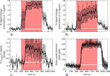
          Blood oxygenation level dependent (BOLD) functional magnetic resonance imaging (fMRI) indirectly measures brain activity based on neurovascular coupling, a reporter that limits both the spatial and temporal resolution of the technique as well as the cellular and metabolic specificity. Emerging methods using functional spectroscopy (fMRS) and diffusion-weighted fMRI suggest that metabolic and structural modifications are also taking place in the activated cells. This paper explores an alternative metabolic imaging approach based on Chemical Exchange Saturation Transfer (CEST) to assess potential metabolic changes induced by neuronal stimulation in rat brains at 17.2 T. An optimized CEST-fMRI data acquisition and processing protocol was developed and used to experimentally assess the feasibility of glucoCEST-based fMRI. Images acquired under glucose-sensitizing conditions showed a substantial negative contrast that highlighted the same brain regions as those activated with BOLD-fMRI. We ascribe this novel fMRI contrast to CEST’s ability to monitor changes in the local concentration of glucose, a metabolite closely coupled to neuronal activity. Our findings are in good agreement with literature employing other modalities. The use of CEST-based techniques for fMRI is not limited to glucose detection; other metabolic pathways involved in neuronal activation could be potentially probed. Moreover, being non invasive, it is conceivable that the same approach can be used for human studies.

          Related collections

          Most cited references45

          • Record: found
          • Abstract: found
          • Article: not found

          Using the amide proton signals of intracellular proteins and peptides to detect pH effects in MRI.

          In the past decade, it has become possible to use the nuclear (proton, 1H) signal of the hydrogen atoms in water for noninvasive assessment of functional and physiological parameters with magnetic resonance imaging (MRI). Here we show that it is possible to produce pH-sensitive MRI contrast by exploiting the exchange between the hydrogen atoms of water and the amide hydrogen atoms of endogenous mobile cellular proteins and peptides. Although amide proton concentrations are in the millimolar range, we achieved a detection sensitivity of several percent on the water signal (molar concentration). The pH dependence of the signal was calibrated in situ, using phosphorus spectroscopy to determine pH, and proton exchange spectroscopy to measure the amide proton transfer rate. To show the potential of amide proton transfer (APT) contrast for detecting acute stroke, pH effects were noninvasively imaged in ischemic rat brain. This observation opens the possibility of using intrinsic pH contrast, as well as protein- and/or peptide-content contrast, as diagnostic tools in clinical imaging.
            Bookmark
            • Record: found
            • Abstract: not found
            • Article: not found

            Intrinsic signal changes accompanying sensory stimulation: functional brain mapping with magnetic resonance imaging.

              Bookmark
              • Record: found
              • Abstract: found
              • Article: not found

              Magnetic Resonance Imaging of Glutamate

              Glutamate (Glu) exhibits a pH and concentration dependent chemical exchange saturation transfer effect (CEST) between its -amine group and bulk water, here termed GluCEST. GluCEST asymmetry is observed at ~3 parts per million downfield from bulk water. Following middle cerebral artery occlusion in the rat brain, an approximately 100% elevation of GluCEST in the ipsilateral side compared to the contralateral side was observed, and is predominantly due to pH changes. In a rat brain tumor model with blood brain barrier disruption, intravenous Glu injection resulted in a clear elevation of GluCEST and a comparable increase in the proton magnetic resonance spectroscopy signal of Glu. GluCEST maps from healthy human brain at 7T were also obtained. These results demonstrate the feasibility and potential of GluCEST for mapping relative changes in Glu concentration as well as pH in vivo. Potential contributions from other brain metabolites to the GluCEST effect are also discussed.
                Bookmark

                Author and article information

                Contributors
                luisa.ciobanu@cea.fr
                Journal
                Sci Rep
                Sci Rep
                Scientific Reports
                Nature Publishing Group UK (London )
                2045-2322
                14 March 2019
                14 March 2019
                2019
                : 9
                : 4423
                Affiliations
                [1 ]GRID grid.457334.2, NeuroSpin, Commissariat à l’Energie Atomique et aux Energies Alternatives, Univerisité Paris-Saclay, ; Gif-sur-Yvette, France
                [2 ]ISNI 0000 0004 0604 7563, GRID grid.13992.30, Department of Chemical and Biological Physics, , Weizmann Institute of Science, ; Rehovot, Israel
                Article
                40986
                10.1038/s41598-019-40986-9
                6418181
                30872689
                f1490cc8-1b01-4fa7-95e8-a99e11904d44
                © The Author(s) 2019

                Open Access This article is licensed under a Creative Commons Attribution 4.0 International License, which permits use, sharing, adaptation, distribution and reproduction in any medium or format, as long as you give appropriate credit to the original author(s) and the source, provide a link to the Creative Commons license, and indicate if changes were made. The images or other third party material in this article are included in the article’s Creative Commons license, unless indicated otherwise in a credit line to the material. If material is not included in the article’s Creative Commons license and your intended use is not permitted by statutory regulation or exceeds the permitted use, you will need to obtain permission directly from the copyright holder. To view a copy of this license, visit http://creativecommons.org/licenses/by/4.0/.

                History
                : 13 April 2018
                : 27 February 2019
                Funding
                Funded by: FundRef https://doi.org/10.13039/501100006489, Commissariat à l'Énergie Atomique et aux Énergies Alternatives (French Alternative Energies and Atomic Energy Commission);
                Funded by: Kimmel Institute for Magnetic Resonance Perlman Family Foundation
                Funded by: Louis-Jeantet Foundation
                Funded by: FundRef https://doi.org/10.13039/501100001665, Agence Nationale de la Recherche (French National Research Agency);
                Award ID: ANR-13-BSV5-0014
                Award Recipient :
                Categories
                Article
                Custom metadata
                © The Author(s) 2019

                Uncategorized
                Uncategorized

                Comments

                Comment on this article