14
views
0
recommends
+1 Recommend
0 collections
    0
    shares
      • Record: found
      • Abstract: found
      • Article: found
      Is Open Access

      Prenatal immune programming of the sex-dependent risk for major depression

      research-article

      Read this article at

      Bookmark
          There is no author summary for this article yet. Authors can add summaries to their articles on ScienceOpen to make them more accessible to a non-specialist audience.

          Abstract

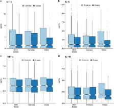
          Maternal immune functioning during pregnancy contributes to sex-dependent deficits in neurodevelopment and to behaviors associated with affective traits in preclinical studies, and has been indirectly associated with offspring depression in epidemiologic studies. We therefore investigated the association between immune activity during pregnancy and the risk of depression among male and female offspring. We conducted a case–control study of depression ( n=484 cases and n=774 controls) using data from the New England Family Study, a pregnancy cohort enrolled between 1959 and 1966 that assessed psychiatric outcomes in adult offspring (mean age=39.7 years). We assayed concentrations of three pro-inflammatory cytokines, interleukin (IL)-1β, IL-6 and tumor necrosis factor (TNF)-α, and the anti-inflammatory cytokine, IL-10, in maternal serum collected at the end of the second and beginning of the third trimesters. High maternal TNF-α was associated with reduced odds of depression among both male and female offspring (odds ratio (OR)=0.68; confidence interval (CI)=0.48, 0.98). However, when considering the TNF-α to IL-10 ratio, a measure of the ratio of pro- to anti-inflammatory loading, maternal immune effects on offspring depression differed significantly by sex ( χ 2=13.9, degrees of freedom=4, P=0.008). Among females, higher maternal TNF-α:IL-10 was associated with reduced odds of depression (OR=0.51; CI=0.32, 0.81), whereas, among males, high maternal TNF-α:IL-10 was associated with elevated odds of depression (OR=1.86; CI=1.02, 3.39). Thus, the balance between TNF-α and IL-10 in maternal prenatal serum was associated with depression in a sex-dependent manner. These findings are consistent with the role of TNF-α in the maturation of the sexually dimorphic fetal brain circuitry that regulates stress and affective responses, and support a prenatal stress-immune model of depression pathogenesis.

          Related collections

          Most cited references40

          • Record: found
          • Abstract: found
          • Article: not found

          Effects of prenatal infection on brain development and behavior: a review of findings from animal models.

          Epidemiological studies with human populations indicate associations between maternal infection during pregnancy and increased risk in offspring for central nervous system (CNS) disorders including schizophrenia, autism and cerebral palsy. Since 2000, a large number of studies have used rodent models of systemic prenatal infection or prenatal immune activation to characterize changes in brain function and behavior caused by the prenatal insult. This review provides a comprehensive summary of these findings, and examines consistencies and trends across studies in an effort to provide a perspective on our current state of understanding from this body of work. Results from these animal modeling studies clearly indicate that prenatal immune activation can cause both acute and lasting changes in behavior and CNS structure and function in offspring. Across laboratories, studies vary with respect to the type, dose and timing of immunogen administration during gestation, species used, postnatal age examined and specific outcome measure quantified. This makes comparison across studies and assessment of replicability difficult. With regard to mechanisms, evidence for roles for several acute mediators of effects of prenatal immune activation has emerged, including circulating interleukin-6, increased placental cytokines and oxidative stress in the fetal brain. However, information required to describe the complete mechanistic pathway responsible for acute effects of prenatal immune activation on fetal brain is lacking, and no studies have yet addressed the issue of how acute prenatal exposure to an immunogen is transduced into a long-term CNS change in the postnatal animal. Directions for further research are discussed. Copyright 2010 Elsevier Inc. All rights reserved.
            Bookmark
            • Record: found
            • Abstract: found
            • Article: not found

            Normal sexual dimorphism of the adult human brain assessed by in vivo magnetic resonance imaging.

            The etiology and consistency of findings on normal sexual dimorphisms of the adult human brain are unresolved. In this study, we present a comprehensive evaluation of normal sexual dimorphisms of cortical and subcortical brain regions, using in vivo magnetic resonance imaging, in a community sample of 48 normal adults. The men and women were similar in age, education, ethnicity, socioeconomic status, general intelligence and handedness. Forty-five brain regions were assessed based on T(1)-weighted three-dimensional images acquired from a 1.5 T magnet. Sexual dimorphisms of adult brain volumes were more evident in the cortex, with women having larger volumes, relative to cerebrum size, particularly in frontal and medial paralimbic cortices. Men had larger volumes, relative to cerebrum size, in frontomedial cortex, the amygdala and hypothalamus. A permutation test showed that, compared to other brain areas assessed in this study, there was greater sexual dimorphism among brain areas that are homologous with those identified in animal studies showing greater levels of sex steroid receptors during critical periods of brain development. These findings have implications for developmental studies that would directly test hypotheses about mechanisms relating sex steroid hormones to sexual dimorphisms in humans.
              Bookmark
              • Record: found
              • Abstract: found
              • Article: not found

              Gonadal steroid hormone receptors and sex differences in the hypothalamo-pituitary-adrenal axis.

              The rapid activation of stress-responsive neuroendocrine systems is a basic reaction of animals to perturbations in their environment. One well-established response is that of the hypothalamo-pituitary-adrenal (HPA) axis. In rats, corticosterone is the major adrenal steroid secreted and is released in direct response to adrenocorticotropin (ACTH) secreted from the anterior pituitary gland. ACTH in turn is regulated by the hypothalamic factor, corticotropin-releasing hormone. A sex difference exists in the response of the HPA axis to stress, with females reacting more robustly than males. It has been demonstrated that in both sexes, products of the HPA axis inhibit reproductive function. Conversely, the sex differences in HPA function are in part due to differences in the circulating gonadal steroid hormone milieu. It appears that testosterone can act to inhibit HPA function, whereas estrogen can enhance HPA function. One mechanism by which androgens and estrogens modulate stress responses is through the binding to their cognate receptors in the central nervous system. The distribution and regulation of androgen and estrogen receptors within the CNS suggest possible sites and mechanisms by which gonadal steroid hormones can influence stress responses. In the case of androgens, data suggest that the control of the hypothalamic paraventricular nucleus is mediated trans-synaptically. For estrogen, modulation of the HPA axis may be due to changes in glucocorticoid receptor-mediated negative feedback mechanisms. The results of a variety of studies suggest that gonadal steroid hormones, particularly testosterone, modulate HPA activity in an attempt to prevent the deleterious effects of HPA activation on reproductive function.
                Bookmark

                Author and article information

                Journal
                Transl Psychiatry
                Transl Psychiatry
                Translational Psychiatry
                Nature Publishing Group
                2158-3188
                May 2016
                31 May 2016
                1 May 2016
                : 6
                : 5
                : e822
                Affiliations
                [1 ]Health Behavior Branch, Division of Intramural Population Health Research, Eunice Kennedy Shriver National Institute of Child Health and Human Development , Rockville, MD, USA
                [2 ]Department of Social and Behavioral Sciences, Harvard T. H. Chan School of Public Health , Boston, MA, USA
                [3 ]Department of Epidemiology, Harvard T. H. Chan School of Public Health , Boston, MA, USA
                [4 ]Department of Psychiatry, Massachusetts General Hospital , Boston, MA, USA
                [5 ]Connors Center for Women’s Health and Gender Biology , Boston, MA, USA
                [6 ]Departments of Psychiatry and Medicine, Harvard Medical School , Boston, MA, USA
                [7 ]Department of Epidemiology, Brown University School of Public Health , Providence, RI, USA
                [8 ]Center for Infection and Immunity, Columbia University Mailman School of Public Health , New York, NY, USA
                [9 ]Department of Epidemiology, Columbia University Mailman School of Public Health, New York, NY, USA
                [10 ]Division of Psychiatric Neuroscience, Massachusetts General Hospital , Boston, MA, USA
                Author notes
                [* ]Health Behavior Branch, Division of Intramural Population Health Research, Eunice Kennedy Shriver National Institute of Child Health and Human Development , 6100 Executive Boulevard, Room 7B13M, Rockville, MD 20852, USA. E-mail stephen.gilman@ 123456nih.gov
                Article
                tp201691
                10.1038/tp.2016.91
                5545649
                27244231
                f19ca1ff-113b-4b05-9edb-35a85d8f23a5
                Copyright © 2016 Macmillan Publishers Limited

                This work is licensed under a Creative Commons Attribution 4.0 International License. The images or other third party material in this article are included in the article’s Creative Commons license, unless indicated otherwise in the credit line; if the material is not included under the Creative Commons license, users will need to obtain permission from the license holder to reproduce the material. To view a copy of this license, visit http://creativecommons.org/licenses/by/4.0/

                History
                : 18 August 2015
                : 23 December 2015
                : 14 January 2016
                Categories
                Original Article

                Clinical Psychology & Psychiatry
                Clinical Psychology & Psychiatry

                Comments

                Comment on this article