Inviting an author to review:
Find an author and click ‘Invite to review selected article’ near their name.
Search for authorsSearch for similar articles
19
views
0
recommends
+1 Recommend
0 collections
    0
    shares
      • Record: found
      • Abstract: found
      • Article: found
      Is Open Access

      Nonfamilial cherubism: A case report and review of literature

      case-report

      Read this article at

      Bookmark
          There is no author summary for this article yet. Authors can add summaries to their articles on ScienceOpen to make them more accessible to a non-specialist audience.

          Abstract

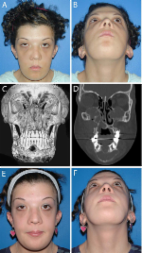
          Cherubism is a rare hereditary developmental condition of the jaws and generally inherited as an autosomal dominant trait. It is also known as familial fibrous dysplasia of the jaws, familial multilocular cystic disease and hereditary fibrous dysplasia of the jaws. The gene for cherubism is mapped to chromosome 4p16.3 may lead to pathologic activation of osteoclasts and disruption of jaw morphogenesis. The lesion usually appears between 2 and 5 years shows a predilection for the mandible and causes a bilateral swelling giving rise to a cherubic chubby appearance. The eosinophilic cuffing of blood vessels appears to be specific for cherubism. The diagnosis is based on clinical, radiographic and histopathologic findings. The purpose of this article is to present a rare case of nonfamilial cherubism as there are very few cases reported and to review the literature with its cone beam computed tomography findings.

          Related collections

          Most cited references22

          • Record: found
          • Abstract: found
          • Article: found
          Is Open Access

          Cherubism: best clinical practice

          Cherubism is a skeletal dysplasia characterized by bilateral and symmetric fibro-osseous lesions limited to the mandible and maxilla. In most patients, cherubism is due to dominant mutations in the SH3BP2 gene on chromosome 4p16.3. Affected children appear normal at birth. Swelling of the jaws usually appears between 2 and 7 years of age, after which, lesions proliferate and increase in size until puberty. The lesions subsequently begin to regress, fill with bone and remodel until age 30, when they are frequently not detectable. Fibro-osseous lesions, including those in cherubism have been classified as quiescent, non-aggressive and aggressive on the basis of clinical behavior and radiographic findings. Quiescent cherubic lesions are usually seen in older patients and do not demonstrate progressive growth. Non-aggressive lesions are most frequently present in teenagers. Lesions in the aggressive form of cherubism occur in young children and are large, rapidly growing and may cause tooth displacement, root resorption, thinning and perforation of cortical bone. Because cherubism is usually self-limiting, operative treatment may not be necessary. Longitudinal observation and follow-up is the initial management in most cases. Surgical intervention with curettage, contouring or resection may be indicated for functional or aesthetic reasons. Surgical procedures are usually performed when the disease becomes quiescent. Aggressive lesions that cause severe functional problems such as airway obstruction justify early surgical intervention.
            Bookmark
            • Record: found
            • Abstract: found
            • Article: not found

            Cherubism - new hypotheses on pathogenesis and therapeutic consequences.

            The hereditary occurrence of cherubism indicates a probable genetic aetiology: a correlation with a mutation in the gene SH3BP2 has been demonstrated. A convincing concept of formal pathogenesis is not yet available. The study was aimed at advancing the understanding of the pathogenesis of cherubism by presenting a case study including genetic findings and an evaluation of the literature. Because of its association with the development of the second and third molars, cherubism could be defined as a genetically determined alteration of tooth development. In this context, disturbed PTHrP - PTHrP receptor interaction induced by the mutation in SH3BP2 is discussed. The temporal and spatial determination of the clinical symptoms is explained by an interaction of SH3BP2-dependent signal transduction pathways with jaw morphogenesis (e.g. Hox-gene Msx-1). Because of the disease-induced lack of determination of the cap phase of the second and third molar, a spatial compartmentation, which is necessary for normal dental development, does not take place. This leads to dysregulation of mesenchymal bone building tissue areas, and to the development of giant cell granulomas with high osteoclastic activity. Because of the genetic determination of cherubism and the associated dedifferentiation of the diseased tissue, a surgical removal should be exclusively restricted to specific indications. Therefore an attitude of wait and see is preferred.
              Bookmark
              • Record: found
              • Abstract: found
              • Article: not found

              Imaging characteristics of cherubism.

              We sought to describe the radiographic and imaging features of cherubism. Cherubism is a rare osseous disorder of children and adolescents. Although the radiologic characteristics of cherubism are not pathognomonic, the diagnosis is strongly suggested by bilateral relatively symmetric jaw involvement that is limited to the maxilla and mandible. Imaging typically shows expansile remodeling of the involved bones, thinning of the cortexes, and multilocular radiolucencies with a coarse trabecular pattern.
                Bookmark

                Author and article information

                Journal
                J Oral Maxillofac Pathol
                J Oral Maxillofac Pathol
                JOMFP
                Journal of Oral and Maxillofacial Pathology : JOMFP
                Medknow Publications & Media Pvt Ltd (India )
                0973-029X
                1998-393X
                Jan-Apr 2017
                : 21
                : 1
                : 181
                Affiliations
                [1]Department of Oral Pathology, Bharati Vidyapeeth Deemed University, Dental College and Hospital, Pune, Maharashtra, India
                [1 ]Department of Oral and Maxillofacial Surgery, Bharati Vidyapeeth Deemed University, Dental College and Hospital, Pune, Maharashtra, India
                Author notes
                Address for correspondence: Dr. Revati Deshmukh, Devikrupa, Shri Dashabhuja Ganesh Cooperative Housing Society, Near Dashabhuja Ganpati Karve Road, Paud Phata, Pune - 411 038, Maharashtra, India. E-mail: rvt_deshmukh@ 123456yahoo.com
                Article
                JOMFP-21-181a
                10.4103/0973-029X.203791
                5406808
                f260eb5b-3a89-42e0-a079-d6e9f6198f13
                Copyright: © 2017 Journal of Oral and Maxillofacial Pathology

                This is an open access article distributed under the terms of the Creative Commons Attribution-NonCommercial-ShareAlike 3.0 License, which allows others to remix, tweak, and build upon the work non-commercially, as long as the author is credited and the new creations are licensed under the identical terms.

                History
                : 01 December 2016
                : 06 December 2016
                Categories
                Case Report

                Pathology
                eosinophilic cuffing,familial fibrous dysplasia,floating teeth appearance,multilocular cystic disease

                Comments

                Comment on this article