0
views
0
recommends
+1 Recommend
0 collections
    0
    shares
      • Record: found
      • Abstract: found
      • Article: found
      Is Open Access

      Catalpol promotes articular cartilage repair by enhancing the recruitment of endogenous mesenchymal stem cells

      research-article

      Read this article at

      Bookmark
          There is no author summary for this article yet. Authors can add summaries to their articles on ScienceOpen to make them more accessible to a non-specialist audience.

          Abstract

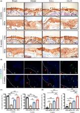
          Articular cartilage defect is challenged by insufficient regenerative ability of cartilage. Catalpol (CA), the primary active component of Rehmanniae Radix, could exert protective effects against various diseases. However, the impact of CA on the treatment of articular cartilage injuries is still unclear. In this study, full‐thickness articular cartilage defect was induced in a mouse model via surgery. The animals were intraperitoneally injected with CA for 4 or 8 weeks. According to the results of macroscopic observation, micro‐computed tomography CT (μCT), histological and immunohistochemistry staining, CA treatment could promote mouse cartilage repair, resulting in cartilage regeneration, bone structure improvement and matrix anabolism. Specifically, an increase in the expression of CD90, the marker of mesenchymal stem cells (MSCs), in the cartilage was observed. In addition, we evaluated the migratory and chondrogenic effects of CA on MSCs. Different concentration of CA was added to C3H10 T1/2 cells. The results showed that CA enhanced cell migration and chondrogenesis without affecting proliferation. Collectively, our findings indicate that CA may be effective for the treatment of cartilage defects via stimulation of endogenous MSCs.

          Related collections

          Most cited references43

          • Record: found
          • Abstract: found
          • Article: not found

          The OARSI histopathology initiative - recommendations for histological assessments of osteoarthritis in the mouse.

          To describe a histologic scoring system for murine osteoarthritis (OA) that can be applied universally to instability, enzymatic, transgenic and spontaneous OA models. Scientists with expertise in assessing murine OA histopathology reviewed the merits and drawbacks of methods described in the literature. A semi-quantitative scoring system that could reasonably be employed in any basic cartilage histology laboratory was proposed. This scoring system was applied to a set of 10 images of the medial tibial plateau and femoral condyle to yield 20 scores. These images were scored twice by four experienced scorers (CL, SG, MC, TA), with a minimum time interval of 1 week between scores to obtain intra-observer variability. An additional three novice scorers (CR, CL and MM) with no previous experience evaluated the images to determine the ease of use and reproducibility across laboratories. The semi-quantitative scoring system was relatively easy to apply for both experienced and novice scorers and the results had low inter- and intra-scorer variability. The variation in scores across both the experienced and novice scorers was low for both tibia and femur, with the tibia always having greater consistency. The semi-quantitative scoring system recommended here is simple to apply and required no specialized equipment. Scoring of the tibial plateaus was highly reproducible and more consistent than that of the femur due to the much thinner femoral cartilage. This scoring system may be a useful tool for both new and experienced scorers to sensitively evaluate models and OA mechanisms, and also provide a common paradigm for comparative evaluation across the many groups performing these analyses. Copyright © 2010 Osteoarthritis Research Society International. Published by Elsevier Ltd. All rights reserved.
            Bookmark
            • Record: found
            • Abstract: found
            • Article: not found

            International Cartilage Repair Society (ICRS) and Oswestry macroscopic cartilage evaluation scores validated for use in Autologous Chondrocyte Implantation (ACI) and microfracture.

            For young patients with cartilage defects, the emergence of clinically applicable cell therapy for biological joint reconstruction is an appealing prospect. Acceptation of this method as a means of standard care requires proof of being reproducible, having long-lasting mechanical integrity, and having a good clinical outcome. This study evaluates the reliability of the International Cartilage Repair Society (ICRS) score and the Oswestry Arthroscopy Score (OAS) in the assessment of regenerative cartilage repair. A total of 101 macroscopic images of cartilage repair were made during arthroscopy 12 months post-treatment of either Autologous Chondrocyte Implantation (ACI) or microfracture. These images were examined by seven independent observers with differing levels of experience. The ICRS and OAS scores were randomly presented twice at a 4-week interval. All observers stated their predicted outcome according to actual treatment and defect size. ICRS and OAS scores showed both good inter- and intra observer reliability (0.62 and 0.56 for ICRS; 0.73 and 0.65 for OAS, respectively). Internal consistency (Cronbach's alpha) was satisfactory for research purposes (0.79 and 0.74, respectively). Correlation (equivalence concordance) between both scoring systems was excellent (r=0.94). All observers were inconsistent in predicting actual treatment. Test-re test reliability of estimated defect size and its correlation to true defect size were poor. These results were also applicable to the sub-analyses of the experience of the observer and the quality of imaging. The ICRS and OAS are reliable and relevant scores that are now both validated for macroscopic evaluation of cartilage repair as a research tool.
              Bookmark
              • Record: found
              • Abstract: found
              • Article: not found

              Osteoarthritis as a disease of the cartilage pericellular matrix

              Osteoarthritis is a painful joint disease characterized by progressive degeneration of the articular cartilage as well as associated changes to the subchondral bone, synovium, and surrounding joint tissues. While the effects of osteoarthritis on the cartilage extracellular matrix (ECM) have been well recognized, it is now becoming apparent that in many cases, the onset of the disease may be initially reflected in the matrix region immediately surrounding the chondrocytes, termed the pericellular matrix (PCM). Growing evidence suggests that the PCM – which along with the enclosed chondrocytes are termed the “chondron” – acts as a critical transducer or “filter” of biochemical and biomechanical signals for the chondrocyte, serving to help regulate the homeostatic balance of chondrocyte metabolic activity in response to environmental signals. Indeed, it appears that alterations in PCM properties and cell-matrix interactions, secondary to genetic, epigenetic, metabolic, or biomechanical stimuli, could in fact serve as initiating or progressive factors for osteoarthritis. Here, we discuss recent advances in the understanding of the role of the PCM, with an emphasis on the reciprocity of changes that occur in this matrix region with disease, as well as how alterations in PCM properties could serve as a driver of ECM-based diseases such as osteoarthritis. Further study of the structure, function, and composition of the PCM in normal and diseased conditions may provide new insights into the understanding of the pathogenesis of osteoarthritis, and presumably new therapeutic approaches for this disease.
                Bookmark

                Author and article information

                Contributors
                tongpeijian@163.com
                hongtingjin@163.com
                Journal
                J Cell Mol Med
                J Cell Mol Med
                10.1111/(ISSN)1582-4934
                JCMM
                Journal of Cellular and Molecular Medicine
                John Wiley and Sons Inc. (Hoboken )
                1582-1838
                1582-4934
                20 March 2024
                April 2024
                : 28
                : 7 ( doiID: 10.1111/jcmm.v28.7 )
                : e18242
                Affiliations
                [ 1 ] Institute of Orthopaedics and Traumatology of Zhejiang Province The First Affiliated Hospital of Zhejiang Chinese Medical University (Zhejiang Provincial Hospital of Chinese Medicine) Hangzhou Zhejiang China
                [ 2 ] The First College of Clinical Medicine Zhejiang Chinese Medical University Hangzhou China
                [ 3 ] Department of Orthopaedic Surgery The First Affiliated Hospital of Zhejiang Chinese Medical University Hangzhou China
                [ 4 ] Department of Orthopaedic Surgery Tongde Hospital of Zhejiang Province Hangzhou China
                Author notes
                [*] [* ] Correspondence

                Hongting Jin and Peijian Tong, Institute of Orthopaedics and Traumatology of Zhejiang Province, The First Affiliated Hospital of Zhejiang Chinese Medical University (Zhejiang Provincial Hospital of Chinese Medicine), Hangzhou, Zhejiang 310006, China.

                Email: hongtingjin@ 123456163.com and tongpeijian@ 123456163.com

                Author information
                https://orcid.org/0000-0001-8795-0874
                Article
                JCMM18242 JCMM-09-2023-268.R1
                10.1111/jcmm.18242
                10955160
                38509736
                f309b531-4c6e-4c28-b2cc-7118a2b3adea
                © 2024 The Authors. Journal of Cellular and Molecular Medicine published by Foundation for Cellular and Molecular Medicine and John Wiley & Sons Ltd.

                This is an open access article under the terms of the http://creativecommons.org/licenses/by/4.0/ License, which permits use, distribution and reproduction in any medium, provided the original work is properly cited.

                History
                : 27 February 2024
                : 30 September 2023
                : 04 March 2024
                Page count
                Figures: 6, Tables: 0, Pages: 12, Words: 5495
                Funding
                Funded by: National Natural Science Foundation of China , doi 10.13039/501100001809;
                Award ID: 82274280
                Funded by: Natural Science Foundation of Zhejiang Province , doi 10.13039/501100004731;
                Award ID: LR23H270001
                Categories
                Original Article
                Original Articles
                Custom metadata
                2.0
                April 2024
                Converter:WILEY_ML3GV2_TO_JATSPMC version:6.3.9 mode:remove_FC converted:21.03.2024

                Molecular medicine
                articular cartilage,cartilage defect,catalpol,chondrogenesis,stem cell homing
                Molecular medicine
                articular cartilage, cartilage defect, catalpol, chondrogenesis, stem cell homing

                Comments

                Comment on this article