32
views
0
recommends
+1 Recommend
0 collections
    0
    shares
      • Record: found
      • Abstract: found
      • Article: found
      Is Open Access

      Deltoid Ligament and Tibiofibular Syndesmosis Injury in Chronic Lateral Ankle Instability: Magnetic Resonance Imaging Evaluation at 3T and Comparison with Arthroscopy

      research-article

      Read this article at

      Bookmark
          There is no author summary for this article yet. Authors can add summaries to their articles on ScienceOpen to make them more accessible to a non-specialist audience.

          Abstract

          Objective

          To evaluate the prevalence of deltoid ligament and distal tibiofibular syndesmosis injury on 3T magnetic resonance imaging (MRI) in patients with chronic lateral ankle instability (CLAI).

          Materials and Methods

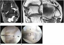
          Fifty patients (mean age, 35 years) who had undergone preoperative 3T MRI and surgical treatment for CLAI were enrolled. The prevalence of deltoid ligament and syndesmosis injury were assessed. The complexity of lateral collateral ligament complex (LCLC) injury was correlated with prevalence of deltoid or syndesmosis injuries. The diagnostic accuracy of ankle ligament imaging at 3T MRI was analyzed using arthroscopy as a reference standard.

          Results

          On MRI, deltoid ligament injury was identified in 18 (36%) patients as follows: superficial ligament alone, 9 (50%); deep ligament alone 2 (11%); and both ligaments 7 (39%). Syndesmosis abnormality was found in 21 (42%) patients as follows: anterior inferior tibiofibular ligament (AITFL) alone, 19 (90%); and AITFL and interosseous ligament, 2 (10%). There was no correlation between LCLC injury complexity and the prevalence of an accompanying deltoid or syndesmosis injury on both MRI and arthroscopic findings. MRI sensitivity and specificity for detection of deltoid ligament injury were 84% and 93.5%, and those for detection of syndesmosis injury were 91% and 100%, respectively.

          Conclusion

          Deltoid ligament or syndesmosis injuries were common in patients undergoing surgery for CLAI, regardless of the LCLC injury complexity. 3T MRI is helpful for the detection of all types of ankle ligament injury. Therefore, careful interpretation of pre-operative MRI is essential.

          Related collections

          Most cited references31

          • Record: found
          • Abstract: found
          • Article: not found

          What is the clinical course of acute ankle sprains? A systematic literature review.

          Ankle sprains are one of the most common musculoskeletal injuries. In order to evaluate the effectiveness of therapeutic interventions and to guide management decisions, it is important to have clear insight of the course of recovery after an acute lateral ankle injury and to evaluate potential factors for nonrecovery and re-sprains. A database search was conducted in MEDLINE, CINAHL, PEDro, EMBASE, and the Cochrane Controlled trial register. Included were observational studies and controlled trials with adult subjects who suffered from an acute lateral ankle sprain that was conventionally treated. One of the following outcomes had to be described: pain, re-sprains, instability, or recovery. Two reviewers independently assessed the methodological quality of each included study. One reviewer extracted relevant data. In total, 31 studies were included, from which 24 studies were of high quality. There was a rapid decrease in pain reporting within the first 2 weeks. Five percent to 33% of patients still experienced pain after 1 year, while 36% to 85% reported full recovery within a period of 3 years. The risk of re-sprains ranged from 3% to 34% of the patients, and re-sprain was registered in periods ranging from 2 weeks to 96 months postinjury. There was a wide variation in subjective instability, ranging from 0% to 33% in the high-quality studies and from 7% to 53% in the low-quality studies. One study described prognostic factors and indicated that training more than 3 times a week is a prognostic factor for residual symptoms. After 1 year of follow-up, a high percentage of patients still experienced pain and subjective instability, while within a period of 3 years, as much as 34% of the patients reported at least 1 re-sprain. From 36% up to 85% of the patients reported full recovery within a period of 3 years.
            Bookmark
            • Record: found
            • Abstract: found
            • Article: not found

            Syndesmosis sprains of the ankle.

            A retrospective review of the clinical records and radiographs of 1344 ankle sprains occurring over a 41-month period at the United States Military Academy was performed. The incidence and severity of these sprains did not differ significantly from other studies with the exception of those injuries involving the tibiofibular ligaments. Those patients sustaining incomplete injuries to the ankle syndesmosis had a recovery time of almost twice that of those patients with severe (3rd degree) ankle sprains (55 vs. 28 days). Eight of 13 patients with 10 of 15 incomplete syndesmosis injuries were available for follow-up examination and radiographs at an average time of 20 months postinjury (range of 7-39 months). All patients with this injury had a positive "squeeze test" (compression of the fibula to the tibia at mid-calf) at the time of initial examination. Nine of 10 patients developed ossification of the syndesmosis in follow-up radiographs. None of these patients had developed chronic ankle instability, loss of motion, or arthritic changes of the joint at the time of final followup.
              Bookmark
              • Record: found
              • Abstract: found
              • Article: not found

              Arthroscopic findings in patients with chronic ankle instability.

              There are little objective data on structural changes of the chronically unstable ankle. Such knowledge could help with preoperative planning. Preoperative ankle arthroscopy provides important insights into the causes and mechanisms of ankle instability and the resulting disability. Case series. From 1993 to 1999, arthroscopic examination was performed in the ankles of 148 patients with symptomatic chronic ankle instability that had lasted 6 months or more. All structural changes were recorded and compared with the clinical diagnosis. A rupture or elongation of the anterior talofibular ligament was noted in 86% of ankles, of the calcaneofibular ligament in 64%, and of the deltoid ligament in 40%. Cartilage damage was noted in 66% of ankles with lateral ligament injuries, whereas 98% of the ankles with deltoid ligament injuries had cartilage damage. Although lateral instability could be verified arthroscopically in 127 patients, medial instability was presumed clinically in 38 patients but was actually detected in 54 patients arthroscopically. Preoperative ankle arthroscopy revealed an essential amount of information that would otherwise have been undetected. For instance, the ligaments showed typical abnormalities corresponding to different entities of ankle instability and different intraarticular pathologic conditions.
                Bookmark

                Author and article information

                Journal
                Korean J Radiol
                Korean J Radiol
                KJR
                Korean Journal of Radiology
                The Korean Society of Radiology
                1229-6929
                2005-8330
                Sep-Oct 2015
                21 August 2015
                : 16
                : 5
                : 1096-1103
                Affiliations
                [1 ]Department of Radiology, Eulji Hospital, Eulji University, Seoul 01830, Korea.
                [2 ]Department of Orthopedic Surgery, Eulji Hospital, Eulji University, Seoul 01830, Korea.
                Author notes
                Corresponding author: Yun Sun Choi, MD, Department of Radiology, Eulji Hospital, Eulji University, 68 Hangeulbiseok-ro, Nowon-gu, Seoul 01830, Korea. Tel: (822) 970-8290, Fax: (822) 970-8346, mdcys0128@ 123456hanmail.net
                Article
                10.3348/kjr.2015.16.5.1096
                4559781
                26356649
                f3725f1e-5405-49ca-8974-4e5b862144c8
                Copyright © 2015 The Korean Society of Radiology

                This is an Open Access article distributed under the terms of the Creative Commons Attribution Non-Commercial License ( http://creativecommons.org/licenses/by-nc/3.0/) which permits unrestricted non-commercial use, distribution, and reproduction in any medium, provided the original work is properly cited.

                History
                : 11 December 2014
                : 02 June 2015
                Categories
                Musculoskeletal Imaging
                Original Article

                Radiology & Imaging
                chronic ankle instability,lateral collateral ligament,deltoid ligament,syndesmosis,magnetic resonance imaging,3t

                Comments

                Comment on this article