0
views
0
recommends
+1 Recommend
0 collections
    0
    shares
      • Record: found
      • Abstract: found
      • Article: found
      Is Open Access

      Deficit Accumulation Index and Biological Markers of Aging in Survivors of Childhood Cancer

      research-article

      Read this article at

      Bookmark
          There is no author summary for this article yet. Authors can add summaries to their articles on ScienceOpen to make them more accessible to a non-specialist audience.

          Abstract

          This cross-sectional study assesses the use of a deficit accumulation index to measure aging-related conditions, such as epigenetic age and telomere length, in adult survivors of acute lymphoblastic leukemia, Hodgkin lymphoma, or central nervous system tumors.

          Key Points

          Question

          Is the deficit accumulation index (DAI), a clinical measure of physiological aging, associated with biomarkers of aging, such as epigenetic age acceleration (EAA) and mean leukocyte telomere length, in survivors of childhood cancer?

          Findings

          In this cross-sectional study of 2101 survivors of childhood cancer, the DAI was associated with EAA but not mean leukocyte telomere length. Both the DAI and EAA were highly effective at identifying aging phenotypes.

          Meaning

          Findings of this study suggest an aging-related biological process underlying the accumulation of deficits among survivors of childhood cancer; either the DAI or EAA may be used to measure aging and response to interventions targeting aging pathways.

          Abstract

          Importance

          Survivors of childhood cancer experience premature aging compared with community controls. The deficit accumulation index (DAI) uses readily available clinical data to measure physiological age in survivors; however, little data exist on how well deficit accumulation represents underlying biological aging among survivors of cancer.

          Objective

          To examine the associations between the DAI and epigenetic age acceleration (EAA) and mean leukocyte telomere length (LTL).

          Design, Setting, and Participants

          This cross-sectional study analyzed data from the St Jude Lifetime Cohort, an assessment of survivors of childhood cancer who were treated at St Jude Children’s Research Hospital in Memphis, Tennessee. Data were collected between 2007 and 2016, assayed between 2014 and 2019, and analyzed between 2022 and 2023. Participants were adult survivors who were diagnosed between 1962 and 2012 and who survived 5 years or more from time of diagnosis. The analyses were restricted to survivors with European ancestry, as there were too few survivors with non-European ancestry.

          Exposures

          The DAI included 44 aging-related items, such as chronic health conditions and functional, psychosocial, and mental well-being. Item responses were summed and divided by the total number of items, resulting in a ratio ranging from 0 to 1. These DAI results were categorized based on reported associations with hospitalization and mortality: low, defined as a DAI less than 0.2; medium, defined as a DAI of 0.2 to less than 0.35; and high, defined as a DAI of 0.35 or higher.

          Main Outcomes and Measures

          Genome-wide DNA methylation was generated from peripheral blood mononuclear cell–derived DNA. The EAA was calculated as the residuals from regressing the Levine epigenetic age on chronological age. The mean LTL was estimated using whole-genome sequencing data.

          Results

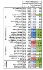
          This study included 2101 survivors of childhood cancer (1122 males [53.4%]; mean [SD] age, 33.9 [9.1] years; median [IQR] time since diagnosis, 25.1 [18.7-31.9] years) with European ancestry. Compared with survivors in the low DAI group, those in the high DAI group experienced 3.7 more years of EAA (β = 3.66; 95% CI, 2.47-4.85; P < .001), whereas those in the medium DAI group experienced 1.8 more years of EAA (β = 1.77; 95% CI, 0.84-2.69; P < .001), independent of treatment exposures. The EAA and DAI association was consistent across 3 common diagnoses (acute lymphoblastic leukemia, Hodgkin lymphoma, and central nervous system tumors) and across chronological age groups. For example, among acute lymphoblastic leukemia survivors, those in the medium DAI group (β = 2.27; 95% CI, 0.78-3.76; P = .001) experienced greater EAA vs those in the low DAI group. Similarly, among survivors younger than 30 years, the high DAI group experienced 4.9 more years of EAA vs the low DAI group (β = 4.95; 95% CI, 2.14-7.75; P < .001). There were no associations between mean LTL residual and the DAI.

          Conclusions and Relevance

          This cross-sectional study of survivors of childhood cancer showed that the DAI was associated with EAA, suggesting an underlying biological process to the accumulation of deficits. Both the DAI and EAA were effective at identifying aging phenotypes, and either may be used to measure aging and response to interventions targeting aging pathways.

          Related collections

          Most cited references38

          • Record: found
          • Abstract: found
          • Article: found
          Is Open Access

          An epigenetic biomarker of aging for lifespan and healthspan

          Identifying reliable biomarkers of aging is a major goal in geroscience. While the first generation of epigenetic biomarkers of aging were developed using chronological age as a surrogate for biological age, we hypothesized that incorporation of composite clinical measures of phenotypic age that capture differences in lifespan and healthspan may identify novel CpGs and facilitate the development of a more powerful epigenetic biomarker of aging. Using an innovative two-step process, we develop a new epigenetic biomarker of aging, DNAm PhenoAge, that strongly outperforms previous measures in regards to predictions for a variety of aging outcomes, including all-cause mortality, cancers, healthspan, physical functioning, and Alzheimer's disease. While this biomarker was developed using data from whole blood, it correlates strongly with age in every tissue and cell tested. Based on an in-depth transcriptional analysis in sorted cells, we find that increased epigenetic, relative to chronological age, is associated with increased activation of pro-inflammatory and interferon pathways, and decreased activation of transcriptional/translational machinery, DNA damage response, and mitochondrial signatures. Overall, this single epigenetic biomarker of aging is able to capture risks for an array of diverse outcomes across multiple tissues and cells, and provide insight into important pathways in aging.
            Bookmark
            • Record: found
            • Abstract: found
            • Article: not found

            DNA methylation-based biomarkers and the epigenetic clock theory of ageing

            Identifying and validating molecular targets of interventions that extend the human health span and lifespan has been difficult, as most clinical biomarkers are not sufficiently representative of the fundamental mechanisms of ageing to serve as their indicators. In a recent breakthrough, biomarkers of ageing based on DNA methylation data have enabled accurate age estimates for any tissue across the entire life course. These 'epigenetic clocks' link developmental and maintenance processes to biological ageing, giving rise to a unified theory of life course. Epigenetic biomarkers may help to address long-standing questions in many fields, including the central question: why do we age?
              Bookmark
              • Record: found
              • Abstract: found
              • Article: found
              Is Open Access

              A standard procedure for creating a frailty index

              Background Frailty can be measured in relation to the accumulation of deficits using a frailty index. A frailty index can be developed from most ageing databases. Our objective is to systematically describe a standard procedure for constructing a frailty index. Methods This is a secondary analysis of the Yale Precipitating Events Project cohort study, based in New Haven CT. Non-disabled people aged 70 years or older (n = 754) were enrolled and re-contacted every 18 months. The database includes variables on function, cognition, co-morbidity, health attitudes and practices and physical performance measures. Data came from the baseline cohort and those available at the first 18-month follow-up assessment. Results Procedures for selecting health variables as candidate deficits were applied to yield 40 deficits. Recoding procedures were applied for categorical, ordinal and interval variables such that they could be mapped to the interval 0–1, where 0 = absence of a deficit, and 1= full expression of the deficit. These individual deficit scores were combined in an index, where 0= no deficit present, and 1= all 40 deficits present. The values of the index were well fit by a gamma distribution. Between the baseline and follow-up cohorts, the age-related slope of deficit accumulation increased from 0.020 (95% confidence interval, 0.014–0.026) to 0.026 (0.020–0.032). The 99% limit to deficit accumulation was 0.6 in the baseline cohort and 0.7 in the follow-up cohort. Multivariate Cox analysis showed the frailty index, age and sex to be significant predictors of mortality. Conclusion A systematic process for creating a frailty index, which relates deficit accumulation to the individual risk of death, showed reproducible properties in the Yale Precipitating Events Project cohort study. This method of quantifying frailty can aid our understanding of frailty-related health characteristics in older adults.
                Bookmark

                Author and article information

                Journal
                JAMA Netw Open
                JAMA Netw Open
                JAMA Network Open
                American Medical Association
                2574-3805
                20 November 2023
                November 2023
                20 November 2023
                : 6
                : 11
                : e2344015
                Affiliations
                [1 ]Department of Epidemiology and Cancer Control, St Jude Children’s Research Hospital, Memphis, Tennessee
                [2 ]Department of Oncology, Georgetown University, Washington, DC
                [3 ]Department of Biostatistics, St Jude Children’s Research Hospital, Memphis, Tennessee
                [4 ]Now with Department of Surgery, Division of Supportive Care in Cancer, University of Rochester School of Medicine and Dentistry, Rochester, New York
                [5 ]Department of Oncology, St Jude Children’s Research Hospital, Memphis, Tennessee
                [6 ]Department of Global Pediatric Medicine, St Jude Children’s Research Hospital, Memphis, Tennessee
                [7 ]Department of Psychology and Biobehavioral Sciences, St Jude Children’s Research Hospital, Memphis, Tennessee
                [8 ]School of Aging Studies, University of South Florida, Tampa
                Author notes
                Article Information
                Accepted for Publication: October 10, 2023.
                Published: November 20, 2023. doi:10.1001/jamanetworkopen.2023.44015
                Open Access: This is an open access article distributed under the terms of the CC-BY License. © 2023 Williams AM et al. JAMA Network Open.
                Corresponding Authors: AnnaLynn Williams, PhD, Department of Surgery, Division of Supportive Care in Cancer, University of Rochester School of Medicine and Dentistry, 265 Crittenden Blvd, Rochester, NY 14642 ( annalynn_williams@ 123456urmc.rochester.edu ); Zhaoming Wang, PhD, Department of Epidemiology and Cancer Control, St Jude Children’s Research Hospital, 262 Danny Thomas Pl, MS 735, Memphis, TN 38105 ( zhaoming.wang@ 123456stjude.org ).
                Author Contributions: Dr Williams had full access to all of the data in the study and takes responsibility for the integrity of the data and the accuracy of the data analysis.
                Concept and design: Williams, Mandelblatt, Armstrong, Brinkman, Robison, Srivastava, Hudson, Ness, Krull, Z. Wang.
                Acquisition, analysis, or interpretation of data: Williams, M. Wang, Dong, Bhakta, Ehrhardt, Mulrooney, Gilmore, Robison, Yasui, Small, Srivastava, Hudson, Ness, Krull, Z. Wang.
                Drafting of the manuscript: Williams, Mandelblatt, M. Wang, Gilmore, Small, Krull, Z. Wang.
                Critical review of the manuscript for important intellectual content: Williams, Mandelblatt, Dong, Armstrong, Bhakta, Brinkman, Ehrhardt, Mulrooney, Gilmore, Robison, Yasui, Small, Srivastava, Hudson, Ness, Krull, Z. Wang.
                Statistical analysis: Williams, M. Wang, Yasui, Small, Srivastava, Z. Wang.
                Obtained funding: Williams, Robison, Hudson, Ness.
                Administrative, technical, or material support: Hudson, Ness, Krull, Z. Wang.
                Supervision: Mandelblatt, Armstrong, Krull, Z. Wang.
                Conflict of Interest Disclosures: Dr Williams reported receiving grants from National Cancer Institute (NCI) at the National Institutes of Health (NIH) during the conduct of the study. Dr Mandelblatt reported receiving grants from NCI/NIH during the conduct of the study. Dr Armstrong reported receiving grants from St Jude Children’s Research Hospital during the conduct of the study. Dr Mulrooney reported receiving grants from NCI/NIH during the conduct of the study. Dr Robison reported receiving grants from NCI/NIH during the conduct of the study. Dr Small reported receiving grants from NCI and grants from National Institute on Aging outside the submitted work. Dr Hudson reported receiving grants from NCI/NIH during the conduct of the study. Dr Ness reported receiving grants from NCI/NIH during the conduct of the study. Dr Krull reported receiving grants from NCI/NIH during the conduct of the study. No other disclosures were reported.
                Funding/Support: This work was supported by grants K00CA222742 and K99CA256356 (Drs Williams, Krull, and Robison), grant U01CA195547 (Drs Hudson and Ness), grant R35CA197289 (Dr Mandelblatt), and grant P30CA021765 from NCI/NIH and by grants from the American Lebanese Syrian Associated Charities.
                Role of the Funder/Sponsor: The funders had no role in the design and conduct of the study; collection, management, analysis, and interpretation of the data; preparation, review, or approval of the manuscript; and decision to submit the manuscript for publication.
                Data Sharing Statement: See Supplement 2.
                Article
                zoi231281
                10.1001/jamanetworkopen.2023.44015
                10660189
                37983031
                f37e4d84-6692-4211-aebf-516c93368544
                Copyright 2023 Williams AM et al. JAMA Network Open.

                This is an open access article distributed under the terms of the CC-BY License.

                History
                : 10 July 2023
                : 10 October 2023
                Categories
                Research
                Original Investigation
                Online Only
                Oncology

                Comments

                Comment on this article