26
views
0
recommends
+1 Recommend
0 collections
    0
    shares
      • Record: found
      • Abstract: found
      • Article: found
      Is Open Access

      Awake craniotomies for aneurysms, arteriovenous malformations, skull base tumors, high flow bypass, and brain stem lesions

      editorial
      Journal of Craniovertebral Junction & Spine
      Medknow Publications & Media Pvt Ltd

      Read this article at

      Bookmark
          There is no author summary for this article yet. Authors can add summaries to their articles on ScienceOpen to make them more accessible to a non-specialist audience.

          Abstract

          Craniotomies for glioma resection under conscious sedation (CS) have been well-documented in the literature for gliomas that are in or adjacent to eloquent areas.[1 2 3 4 5] To the best of our knowledge, based on a review of current literature, the use of awake surgery forclipping of aneurysms, high flow extracranial to intercranial (EC-IC) bypass, resection of arteriovenous malformations (AVMs), resection of skull base tumors, and the resection of brain stem lesions has not previously been reported. Intraoperative monitoring using electroencephalography (EEG), somatosensory evoked potentials (SSEP), and motor evoked potentials (MEP) have inherent false-positives and-negatives that have been reported and have been experienced by neurosurgeons during craniotomies for the abovementioned pathologies. During cerebral aneurysm surgery, temporary clipping is often required, and by proposing craniotomy under CS, we are capable of assessing neurological function (i.e., hand-motor function during middle cerebral artery division temporary clipping). At the final reconstruction of the aneurysm and by assessing neurological function, we are able to evaluate any potential risk to a perforator behind the aneurysm by performing a direct neurological evaluation. During microsurgical resection ofAVMs located in or around eloquent areas can be challenging. This is due, in large part, to the risk of obliterating vessels in the area of the AVM nidus that may be supplying normal cortical and subcortical structures. By checking each one of these feeder versus bystander vessel by performing direct neurological examination, we are able to avoid the risk of ischemia to normal brain tissue. During high-flow EC-IC bypass surgery, two important steps, temporary occlusion of the recipient vessel and the permanent occlusion of the parent aneurysmal vessel, are monitored using electrophysiology, which can have both false-positives and -negatives. The risk of the latter can be significantly avoided if the procedure can be performed in a patient under conscious ("awake") sedation, allowing direct motor evaluation. During brain stem surgery, monitoring technology is even more limited. By performing the procedure under CS, we are able to test each function related to the respective region being operated on and thus potentially decreasing the neurological morbidity of the procedure. Anesthesia team protocol is as follows: The surgical procedure is performed under a monitored anesthesia care. Spontaneous respirations are maintained throughout the case with dexmedetomidine (0.2-1.0 mcg/kg/h) and remifentanil (0.05-2 mcg/kg/min) infusions. EEG monitoring is used to help gauge the appropriate depth of anesthesia. A scalp block using 0.5% ropivicaine with epinephrine is used to anesthetize the respective scalp region. Prior to the awake-phase of the surgery, thedexmedetomidineinfusion is discontinued. Remifentanil is continued for pain control, and elevations in blood pressure are treated with nitroglycerin and esmolol. Neurological evaluation is then focused on the function of interest depending on the specific procedure. Sedation is then restarted for the remainder of the surgery. Motor function is tested for the respective region by a member of the neurosurgery 
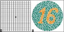
team. Vision testing is performed by using color plates and an iPad with specific programs quantifying various visual tests [Figures 1 and 2]. Motor cranial nerve function is tested directly. Figure 1 Intraoperative photograph taken during temporary clipping of vessels surrounding a pulvinar-occipital (arteriovenous malformation) AVM Figure 2 (a) Amsler grid and (b) color plate We have now performed 61 craniotomies using the above awake/CS protocol. No single case had to be converted into general anesthesia during or after the procedure. Compared to our own series prior to the awake procedures, for the same cases, we have lower neurological morbidity, zero mortality, and shorter hospital stay in this cohort.

          Related collections

          Most cited references4

          • Record: found
          • Abstract: found
          • Article: not found

          Awake mapping optimizes the extent of resection for low-grade gliomas in eloquent areas.

          Awake craniotomy with intraoperative electrical mapping is a reliable method to minimize the risk of permanent deficit during surgery for low-grade glioma located within eloquent areas classically considered inoperable. However, it could be argued that preservation of functional sites might lead to a lesser degree of tumor removal. To the best of our knowledge, the extent of resection has never been directly compared between traditional and awake procedures. We report for the first time a series of patients who underwent 2 consecutive surgeries without and with awake mapping. Nine patients underwent surgery for a low-grade glioma in functional sites under general anesthesia in other institutions. The resection was subtotal in 3 cases and partial in 6 cases. There was a postoperative worsening in 3 cases. We performed a second surgery in the awake condition with intraoperative electrostimulation. The resection was performed according to functional boundaries at both the cortical and subcortical levels. Postoperative magnetic resonance imaging showed that the resection was complete in 5 cases and subtotal in 4 cases (no partial removal) and that it was improved in all cases compared with the first surgery (P = .04). There was no permanent neurological worsening. Three patients improved compared with the presurgical status. All patients returned to normal professional and social lives. Our results demonstrate that awake surgery, known to preserve the quality of life in patients with low-grade glioma, is also able to significantly improve the extent of resection for lesions located in functional regions.
            Bookmark
            • Record: found
            • Abstract: found
            • Article: not found

            Awake craniotomy with brain mapping as the routine surgical approach to treating patients with supratentorial intraaxial tumors: a prospective trial of 200 cases.

            Awake craniotomy was performed as the standard surgical approach to supratentorial intraaxial tumors, regardless of the involvement of eloquent cortex, in a prospective trial of 200 patients surgically treated by the same surgeon at a single institution. Patient presentations, comorbid conditions, tumor locations, and the histological characteristics of lesions were recorded. Brain mapping was possible in 195 (97.5%) of 200 patients. The total number of patients sustaining complications was 33 for an overall complication rate of 16.5%. There were two deaths in this series, for a mortality rate of 1%. New postoperative neurological deficits were seen in 13% of the patients, but these were permanent in only 4.5% of them. Complication rates were higher in patients who had gliomas or preoperative neurological deficits and in those who had undergone prior radiation therapy or surgery. No patient who entered the operating room neurologically intact sustained a permanent neurological deficit postoperatively. Of the most recent 50 patients treated, three (6%) required a stay in the intensive care unit, and the median total hospital stay was 1 day. Use of awake craniotomy can result in a considerable reduction in resource utilization without compromising patient care by minimizing intensive care time and total hospital stay. Awake craniotomy is a practical and effective standard surgical approach to supratentorial tumors with a low complication rate, and provides an excellent alternative to craniotomy performed with the patient in the state of general anesthesia because it allows the opportunity for brain mapping and avoids general anesthesia.
              Bookmark
              • Record: found
              • Abstract: found
              • Article: not found

              The predictive value of intraoperative somatosensory evoked potential monitoring: review of 244 procedures.

              There is some controversy regarding the value of intraoperative neurophysiological monitoring in predicting postoperative neurological deficits. We discuss our experience with the use of intraoperative somatosensory evoked potentials (SSEPs) during surgery of cranial base tumors. We retrospectively reviewed all of the procedures that had been performed for the resection of cranial base tumors from July 29, 1993, through March 16, 1995. One hundred ninety-three consecutive patients had undergone a total of 244 procedures. SSEP waveforms were classified as follows: Type I, no change; Type II, change that reverts to baseline; Type III, change that does not revert to baseline; and Type IV, complete flattening of the SSEP waveform without improvement. Two patients had no waveforms from the beginning of the case (Type V) and were excluded from further analysis. New immediate postoperative neurological deficits were recorded. There were 64 male and 129 female patients, with a mean age of 46.6 years. One hundred seventy-seven patients had Type I SSEP waveforms, 13 of whom had postoperative deficits (7%). Fifty-six patients had Type II SSEPs, and nine (16%) of them had postoperative neurological deficits. Six patients had Type III SSEPs, and three had Type IV SSEPs, all of whom (100%) had postoperative deficits. There was a correlation between SSEP type and the results of the postoperative neurological examinations. The positive predictive value is 100%, and the negative predictive value is 90%. Although a change in the waveform that did not revert to baseline (Types III and IV) always predicted a postoperative deficit, a normal waveform did not always rule out postoperative deficits. Pathological abnormality, vessel encasement, vessel narrowing, degree of cavernous sinus involvement, brain stem edema, middle fossa location, final amount of resection, age, and tumor size correlated with a high predictive value of SSEP monitoring on univariate analysis (P 0.05), although brain stem edema was close (P = 0.0571). Intraoperative SSEPs have a high positive predictive value during surgery for cranial base tumors, but they do not detect all postoperative deficits.
                Bookmark

                Author and article information

                Journal
                J Craniovertebr Junction Spine
                J Craniovertebr Junction Spine
                JCVJS
                Journal of Craniovertebral Junction & Spine
                Medknow Publications & Media Pvt Ltd (India )
                0974-8237
                0976-9285
                Jan-Mar 2015
                : 6
                : 1
                : 8-9
                Affiliations
                [1]Department of Neurosurgery, Saint Louis University, Saint Louis, Missouri, USA
                Author notes
                Corresponding author: Prof. Saleem I. Abdulrauf, 3635 Vista Avenue, Saint Louis - 63110, Missour, United States. E-mail: abdulrsi@ 123456slu.edu
                Article
                JCVJS-6-8
                10.4103/0974-8237.151580
                4361840
                25788813
                f395d375-6f77-4fa6-a3a8-189416131670
                Copyright: © Journal of Craniovertebral Junction and Spine

                This is an open-access article distributed under the terms of the Creative Commons Attribution-Noncommercial-Share Alike 3.0 Unported, which permits unrestricted use, distribution, and reproduction in any medium, provided the original work is properly cited.

                History
                Categories
                Editorial

                Neurology
                Neurology

                Comments

                Comment on this article