0
views
0
recommends
+1 Recommend
0 collections
    0
    shares
      • Record: found
      • Abstract: found
      • Article: found
      Is Open Access

      In Situ Glycan Analysis and Editing in Living Systems

      review-article

      Read this article at

      Bookmark
          There is no author summary for this article yet. Authors can add summaries to their articles on ScienceOpen to make them more accessible to a non-specialist audience.

          Abstract

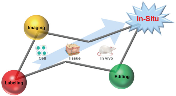
          Besides proteins and nucleic acids, carbohydrates are also ubiquitous building blocks of living systems. Approximately 70% of mammalian proteins are glycosylated. Glycans not only provide structural support for living systems but also act as crucial regulators of cellular functions. As a result, they are considered essential pieces of the life science puzzle. However, research on glycans has lagged far behind that on proteins and nucleic acids. The main reason is that glycans are not direct products of gene coding, and their synthesis is nontemplated. In addition, the diversity of monosaccharide species and their linkage patterns contribute to the complexity of the glycan structures, which is the molecular basis for their diverse functions. Research in glycobiology is extremely challenging, especially for the in situ elucidation of glycan structures and functions. There is an urgent need to develop highly specific glycan labeling tools and imaging methods and devise glycan editing strategies. This Perspective focuses on the challenges of in situ analysis of glycans in living systems at three spatial levels (i.e., cell, tissue, and in vivo) and highlights recent advances and directions in glycan labeling, imaging, and editing tools. We believe that examining the current development landscape and the existing bottlenecks can drive the evolution of in situ glycan analysis and intervention strategies and provide glycan-based insights for clinical diagnosis and therapeutics.

          Related collections

          Most cited references205

          • Record: found
          • Abstract: found
          • Article: not found

          Glycosylation in cellular mechanisms of health and disease.

          Glycosylation produces an abundant, diverse, and highly regulated repertoire of cellular glycans that are frequently attached to proteins and lipids. The past decade of research on glycan function has revealed that the enzymes responsible for glycosylation-the glycosyltransferases and glycosidases-are essential in the development and physiology of living organisms. Glycans participate in many key biological processes including cell adhesion, molecular trafficking and clearance, receptor activation, signal transduction, and endocytosis. This review discusses the increasingly sophisticated molecular mechanisms being discovered by which mammalian glycosylation governs physiology and contributes to disease.
            Bookmark
            • Record: found
            • Abstract: found
            • Article: not found

            Complex N-glycan number and degree of branching cooperate to regulate cell proliferation and differentiation.

            The number of N-glycans (n) is a distinct feature of each glycoprotein sequence and cooperates with the physical properties of the Golgi N-glycan-branching pathway to regulate surface glycoprotein levels. The Golgi pathway is ultrasensitive to hexosamine flux for the production of tri- and tetra-antennary N-glycans, which bind to galectins and form a molecular lattice that opposes glycoprotein endocytosis. Glycoproteins with few N-glycans (e.g., TbetaR, CTLA-4, and GLUT4) exhibit enhanced cell-surface expression with switch-like responses to increasing hexosamine concentration, whereas glycoproteins with high numbers of N-glycans (e.g., EGFR, IGFR, FGFR, and PDGFR) exhibit hyperbolic responses. Computational and experimental data reveal that these features allow nutrient flux stimulated by growth-promoting high-n receptors to drive arrest/differentiation programs by increasing surface levels of low-n glycoproteins. We have identified a mechanism for metabolic regulation of cellular transition between growth and arrest in mammals arising from apparent coevolution of N-glycan number and branching.
              Bookmark
              • Record: found
              • Abstract: found
              • Article: not found

              Glycosylation and the immune system.

              Almost all of the key molecules involved in the innate and adaptive immune response are glycoproteins. In the cellular immune system, specific glycoforms are involved in the folding, quality control, and assembly of peptide-loaded major histocompatibility complex (MHC) antigens and the T cell receptor complex. Although some glycopeptide antigens are presented by the MHC, the generation of peptide antigens from glycoproteins may require enzymatic removal of sugars before the protein can be cleaved. Oligosaccharides attached to glycoproteins in the junction between T cells and antigen-presenting cells help to orient binding faces, provide protease protection, and restrict nonspecific lateral protein-protein interactions. In the humoral immune system, all of the immunoglobulins and most of the complement components are glycosylated. Although a major function for sugars is to contribute to the stability of the proteins to which they are attached, specific glycoforms are involved in recognition events. For example, in rheumatoid arthritis, an autoimmune disease, agalactosylated glycoforms of aggregated immunoglobulin G may induce association with the mannose-binding lectin and contribute to the pathology.
                Bookmark

                Author and article information

                Journal
                JACS Au
                JACS Au
                au
                jaaucr
                JACS Au
                American Chemical Society
                2691-3704
                17 January 2024
                26 February 2024
                : 4
                : 2
                : 384-401
                Affiliations
                []State Key Laboratory of Analytical Chemistry for Life Science, School of Chemistry and Chemical Engineering, Nanjing University , Nanjing 210023, China
                []Chemistry and Biomedicine Innovation Center (ChemBIC), Nanjing University , Nanjing 210023, China
                Author notes
                Author information
                https://orcid.org/0000-0002-3775-3028
                https://orcid.org/0000-0001-5381-3484
                https://orcid.org/0000-0002-6741-5302
                Article
                10.1021/jacsau.3c00717
                10900212
                38425935
                f570479f-e13c-49e5-b68a-2b0d6def748d
                © 2024 The Authors. Published by American Chemical Society

                Permits non-commercial access and re-use, provided that author attribution and integrity are maintained; but does not permit creation of adaptations or other derivative works ( https://creativecommons.org/licenses/by-nc-nd/4.0/).

                History
                : 15 November 2023
                : 19 December 2023
                : 15 December 2023
                Funding
                Funded by: National Natural Science Foundation of China, doi 10.13039/501100001809;
                Award ID: 21827812
                Funded by: Fundamental Research Funds for the Central Universities, doi 10.13039/501100012226;
                Award ID: 2022300324
                Funded by: Fundamental Research Funds for the Central Universities, doi 10.13039/501100012226;
                Award ID: 021414380502
                Funded by: Fundamental Research Funds for the Central Universities, doi 10.13039/501100012226;
                Award ID: 020514380309
                Funded by: State Key Laboratory of Analytical Chemistry for Life Sciences, doi 10.13039/501100011244;
                Award ID: SKLACLS2104
                Funded by: State Key Laboratory of Analytical Chemistry for Life Sciences, doi 10.13039/501100011244;
                Award ID: 5431ZZXM2305
                Funded by: State Key Laboratory of Analytical Chemistry for Life Sciences, doi 10.13039/501100011244;
                Award ID: 5431ZZXM2204
                Funded by: National Natural Science Foundation of China, doi 10.13039/501100001809;
                Award ID: 22274073
                Funded by: National Natural Science Foundation of China, doi 10.13039/501100001809;
                Award ID: 21974067
                Funded by: National Natural Science Foundation of China, doi 10.13039/501100001809;
                Award ID: 21890741
                Categories
                Perspective
                Custom metadata
                au3c00717
                au3c00717

                glycan,living system,in situ,glycan labeling,glycan imaging,glycan editing

                Comments

                Comment on this article