0
views
0
recommends
+1 Recommend
0 collections
    0
    shares
      • Record: found
      • Abstract: found
      • Article: found
      Is Open Access

      Analysis of the distribution of vagal afferent projections from different peripheral organs to the nucleus of the solitary tract in rats

      research-article

      Read this article at

      Bookmark
          There is no author summary for this article yet. Authors can add summaries to their articles on ScienceOpen to make them more accessible to a non-specialist audience.

          Abstract

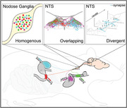
          Anatomical tracing studies examining the vagal system can conflate details of sensory afferent and motor efferent neurons. Here, we used a serotype of adeno‐associated virus that transports retrogradely and exhibits selective tropism for vagal afferents, to map their soma location and central termination sites within the nucleus of the solitary tract (NTS). We examined the vagal sensory afferents innervating the trachea, duodenum, stomach, or heart, and in some animals, from two organs concurrently. We observed no obvious somatotopy in the somata distribution within the nodose ganglion. The central termination patterns of afferents from different organs within the NTS overlap substantially. Convergence of vagal afferent inputs from different organs onto single NTS neurons is observed. Abdominal and thoracic afferents terminate throughout the NTS, including in the rostral NTS, where the 7th cranial nerve inputs are known to synapse. To address whether the axonal labeling produced by viral transduction is so widespread because it fills axons traveling to their targets, and not just terminal fields, we labeled pre and postsynaptic elements of vagal afferents in the NTS . Vagal afferents form multiple putative synapses as they course through the NTS, with each vagal afferent neuron distributing sensory signals to multiple second‐order NTS neurons. We observe little selectivity between vagal afferents from different visceral targets and NTS neurons with common neurochemical phenotypes, with afferents from different organs making close appositions with the same NTS neuron. We conclude that specific viscerosensory information is distributed widely within the NTS and that the coding of this input is probably determined by the intrinsic properties and projections of the second‐order neuron.

          Abstract

          We injected retrograde pseudotyped adeno‐associated viruses into different peripheral organs and mapped the axonal projections of vagal viscerosensory neurons within the nucleus of the solitary tract (NTS). We observed no obvious viscerotopy, with axons from different organs overlapping in NTS subnuclei and with NTS neurons of different neurochemical phenotype. The implications for deciphering this viscerosensory code are discussed.

          Related collections

          Most cited references81

          • Record: found
          • Abstract: found
          • Article: not found

          iDISCO: a simple, rapid method to immunolabel large tissue samples for volume imaging.

          The visualization of molecularly labeled structures within large intact tissues in three dimensions is an area of intense focus. We describe a simple, rapid, and inexpensive method, iDISCO, that permits whole-mount immunolabeling with volume imaging of large cleared samples ranging from perinatal mouse embryos to adult organs, such as brains or kidneys. iDISCO is modeled on classical histology techniques, facilitating translation of section staining assays to intact tissues, as evidenced by compatibility with 28 antibodies to both endogenous antigens and transgenic reporters like GFP. When applied to degenerating neurons, iDISCO revealed unexpected variability in number of apoptotic neurons within individual sensory ganglia despite tight control of total number in all ganglia. It also permitted imaging of single degenerating axons in adult brain and the first visualization of cleaved Caspase-3 in degenerating embryonic sensory axons in vivo, even single axons. iDISCO enables facile volume imaging of immunolabeled structures in complex tissues. PAPERCLIP:
            Bookmark
            • Record: found
            • Abstract: found
            • Article: not found

            A Designer AAV Variant Permits Efficient Retrograde Access to Projection Neurons

            Efficient retrograde access to projection neurons for the delivery of sensors and effectors constitutes an important and enabling capability for neural circuit dissection. Such an approach would also be useful for gene therapy, including the treatment of neurodegenerative disorders characterized by pathological spread through functionally connected and highly distributed networks. Viral vectors, in particular, are powerful gene delivery vehicles for the nervous system, but all available tools suffer from inefficient retrograde transport or limited clinical potential. To address this need, we applied in vivo directed evolution to engineer potent retrograde functionality into the capsid of adeno-associated virus (AAV), a vector that has shown promise in neuroscience research and the clinic. A newly evolved variant, rAAV2-retro, permits robust retrograde access to projection neurons with efficiency comparable to classical synthetic retrograde tracers and enables sufficient sensor/effector expression for functional circuit interrogation and in vivo genome editing in targeted neuronal populations. VIDEO ABSTRACT.
              Bookmark
              • Record: found
              • Abstract: found
              • Article: not found

              Functional and chemical anatomy of the afferent vagal system.

              The results of neural tracing studies suggest that vagal afferent fibers in cervical and thoracic branches innervate the esophagus, lower airways, heart, aorta, and possibly the thymus, and via abdominal branches the entire gastrointestinal tract, liver, portal vein, billiary system, pancreas, but not the spleen. In addition, vagal afferents innervate numerous thoracic and abdominal paraganglia associated with the vagus nerves. Specific terminal structures such as flower basket terminals, intraganglionic laminar endings and intramuscular arrays have been identified in the various organs and organ compartments, suggesting functional specializations. Electrophysiological recording studies have identified mechano- and chemo-receptors, as well as temperature- and osmo-sensors. In the rat and several other species, mostly polymodal units, while in the cat more specialized units have been reported. Few details of the peripheral transduction cascades and the transmitters for signal propagation in the CNS are known. Glutamate and its various receptors are likely to play an important role at the level of primary afferent signaling to the solitary nucleus. The vagal afferent system is thus in an excellent position to detect immune-related events in the periphery and generate appropriate autonomic, endocrine, and behavioral responses via central reflex pathways. There is also good evidence for a role of vagal afferents in nociception, as manifested by affective-emotional responses such as increased blood pressure and tachycardia, typically associated with the perception of pain, and mediated via central reflex pathways involving the amygdala and other parts of the limbic system. The massive central projections are likely to be responsible for the antiepileptic properties of afferent vagal stimulation in humans. Furthermore, these functions are in line with a general defensive character ascribed to the vagal afferent, paraventricular system in lower vertebrates.
                Bookmark

                Author and article information

                Contributors
                stuart.mcdougall@florey.edu.au
                a.allen@unimelb.edu.au
                Journal
                J Comp Neurol
                J Comp Neurol
                10.1002/(ISSN)1096-9861
                CNE
                The Journal of Comparative Neurology
                John Wiley and Sons Inc. (Hoboken )
                0021-9967
                1096-9861
                21 August 2022
                December 2022
                : 530
                : 17 ( doiID: 10.1002/cne.v530.17 )
                : 3072-3103
                Affiliations
                [ 1 ] Department of Anatomy and Physiology The University of Melbourne Parkville Victoria Australia
                [ 2 ] Department of Biomedical Engineering Case Western Reserve University Cleveland Ohio USA
                [ 3 ] Department of Queensland Brain Institute The University of Queensland St Lucia Queensland Australia
                [ 4 ] Department of Florey Institute of Neuroscience and Mental Health The University of Melbourne Parkville Victoria Australia
                Author notes
                [*] [* ] Correspondence

                Stuart J. McDougall, Florey Institute of Neuroscience and Mental Health, The University of Melbourne, Parkville, Victoria 3010, Australia.

                Email: stuart.mcdougall@ 123456florey.edu.au

                Andrew M. Allen, Department of Anatomy and Physiology, The University of Melbourne, Parkville, Victoria 3010, Australia.

                Email: a.allen@ 123456unimelb.edu.au

                Author information
                https://orcid.org/0000-0002-4397-9277
                https://orcid.org/0000-0002-8364-6266
                https://orcid.org/0000-0002-8778-675X
                https://orcid.org/0000-0002-2183-5360
                Article
                CNE25398
                10.1002/cne.25398
                9804483
                35988033
                f5a514f4-91ff-4f33-bee7-7148d7f36531
                © 2022 The Authors. The Journal of Comparative Neurology published by Wiley Periodicals LLC.

                This is an open access article under the terms of the http://creativecommons.org/licenses/by-nc-nd/4.0/ License, which permits use and distribution in any medium, provided the original work is properly cited, the use is non‐commercial and no modifications or adaptations are made.

                History
                : 26 July 2022
                : 21 March 2022
                : 02 August 2022
                Page count
                Figures: 14, Tables: 2, Pages: 32, Words: 20419
                Funding
                Funded by: National Health and Medical Research Council , doi 10.13039/501100000925;
                Award ID: 1156727
                Award ID: 1163039
                Funded by: Australian Research Council , doi 10.13039/501100000923;
                Award ID: DP210102546
                Categories
                Research Article
                Research Articles
                Custom metadata
                2.0
                December 2022
                Converter:WILEY_ML3GV2_TO_JATSPMC version:6.2.3 mode:remove_FC converted:31.12.2022

                Neurology
                viscerosensory afferent,dorsal motor nucleus of vagus,area postrema,stomach,duodenum,heart,trachea

                Comments

                Comment on this article