9
views
0
recommends
+1 Recommend
0 collections
    0
    shares
      • Record: found
      • Abstract: found
      • Article: found
      Is Open Access

      Development of a Novel Nanotextured Titanium Implant. An Experimental Study in Rats

      research-article

      Read this article at

      Bookmark
          There is no author summary for this article yet. Authors can add summaries to their articles on ScienceOpen to make them more accessible to a non-specialist audience.

          Abstract

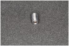
          This animal study evaluated the osseointegration level of a new nanotextured titanium surface produced by anodization. Ti-cp micro-implants (1.5 mm diameter by 2.5 mm in length) divided into two groups: titanium nanotextured surface treatment (Test Group) and acid etched surface treatment (Control Group). Surface characterization included morphology analysis using scanning electron microscopy and wettability by measuring contact angle. Sixteen Wistar rats were submitted to two micro implants surgical placement procedures. In each rat, one type of micro implant placed in each tibia. The animals sacrificed after two (T1) and six weeks (T2) post-implantation. After the euthanasia, tibias processed for histomorphometric analysis, which allowed the evaluation of bone to implant contact (BIC) and the bone area fraction occupancy between the threads (BAFO). Our surface analysis data showed that the Control Group exhibited an irregular and non-homogenous topography while the Test Group showed a nanotextured surface. The Test Group showed higher wettability (contact angle = 5.1 ± 0.7°) than the Control Group (contact angle = 75.5 ± 4.6°). Concerning the histomorphometric analysis results for T1, Control and Test groups showed BIC percentages of 41.3 ± 15.2% and 63.1 ± 8.7% ( p < 0.05), respectively, and for BAFO, 28.7 ± 13.7% and 54.8 ± 7.5%, respectively ( p < 0.05). For T2, the histomorphometric analysis for Control and Test groups showed BIC percentages of 51.2 ± 11.4% and 64.8 ± 7.4% ( p < 0.05), respectively and for BAFO, 36.4 ± 10.3% and 57.9 ± 9.3% ( p < 0.05), respectively. The findings of the current study confirmed that the novel nanotextured surface exhibited superior wettability, improved peri-implant bone formation, and expedited osseointegration.

          Related collections

          Most cited references42

          • Record: found
          • Abstract: found
          • Article: not found

          Influence of surface characteristics on bone integration of titanium implants. A histomorphometric study in miniature pigs.

          The purpose of the present study was to evaluate the influence of different surface characteristics on bone integration of titanium implants. Hollow-cylinder implants with six different surfaces were placed in the metaphyses of the tibia and femur in six miniature pigs. After 3 and 6 weeks, the implants with surrounding bone were removed and analyzed in undecalcified transverse sections. The histologic examination revealed direct bone-implant contact for all implants. However, the morphometric analyses demonstrated significant differences in the percentage of bone-implant contact, when measured in cancellous bone. Electropolished as well as the sandblasted and acid pickled (medium grit; HF/HNO3) implant surfaces had the lowest percentage of bone contact with mean values ranging between 20 and 25%. Sandblasted implants with a large grit and titanium plasma-sprayed implants demonstrated 30-40% mean bone contact. The highest extent of bone-implant interface was observed in sandblasted and acid attacked surfaces (large grit; HCl/H2SO4) with mean values of 50-60%, and hydroxylapatite (HA)-coated implants with 60-70%. However, the HA coating consistently revealed signs of resorption. It can be concluded that the extent of bone-implant interface is positively correlated with an increasing roughness of the implant surface.
            Bookmark
            • Record: found
            • Abstract: found
            • Article: not found

            Titanium surface characteristics, including topography and wettability, alter macrophage activation.

            Biomaterial surface properties including chemistry, topography, and wettability regulate cell response. Previous studies have shown that increasing surface roughness of metallic orthopaedic and dental implants improved bone formation around the implant. Little is known about how implant surface properties can affect immune cells that generate a wound healing microenvironment. The aim of our study was to examine the effect of surface modifications on macrophage activation and cytokine production. Macrophages were cultured on seven surfaces: tissue culture polystyrene (TCPS) control; hydrophobic and hydrophilic smooth Ti (PT and oxygen-plasma-treated (plasma) PT); hydrophobic and hydrophilic microrough Ti (SLA and plasma SLA), and hydrophobic and hydrophilic nano-and micro-rough Ti (aged modSLA and modSLA). Smooth Ti induced inflammatory macrophage (M1-like) activation, as indicated by increased levels of interleukins IL-1β, IL-6, and TNFα. In contrast, hydrophilic rough titanium induced macrophage activation similar to the anti-inflammatory M2-like state, increasing levels of interleukins IL-4 and IL-10. These results demonstrate that macrophages cultured on high surface wettability materials produce an anti-inflammatory microenvironment, and this property may be used to improve the healing response to biomaterials.
              Bookmark
              • Record: found
              • Abstract: found
              • Article: not found

              Early osseointegration to hydrophilic and hydrophobic implant surfaces in humans.

              To evaluate the rate and degree of osseointegration at chemically modified moderately rough, hydrophilic (SLActive) and moderately rough, hydrophobic (SLA) implant surfaces during early phases of healing in a human model. The devices used for this study of early healing were 4 mm long and 2.8 mm in diameter and had either an SLActive chemically modified or a moderately rough SLA surface configuration. These devices were surgically installed into the retro-molar area of 49 human volunteers and retrieved after 7, 14, 28 and 42 days of submerged healing. A 5.2-mm-long specially designed trephine with a 4.9 mm inside diameter, allowing the circumferential sampling of 1 mm tissue together with the device was applied. Histologic ground sections were prepared and histometric analyses of the tissue components (i.e. old bone, new bone, bone debris and soft tissue) in contact with the device surfaces were performed. All device sites healed uneventfully. All device surfaces were partially coated with bone debris. A significant fraction of this bone matrix coating became increasingly covered with newly formed bone. The process of new bone formation started already during the first week in the trabecular regions and increased gradually up to 42 days. The percentage of direct contact between newly formed bone and the device (bone-to-implant contact) after 2 and 4 weeks was more pronounced adjacent to the SLActive than to the SLA surface (14.8% vs. 12.2% and 48.3% vs. 32.4%, respectively), but after 42 days, these differences were no longer evident (61.6% vs. 61.5%). While healing showed similar characteristics with bone resorptive and appositional events for both SLActive and SLA surfaces between 7 and 42 days, the degree of osseointegration after 2 and 4 weeks was superior for the SLActive compared with the SLA surface. © 2011 John Wiley & Sons A/S.
                Bookmark

                Author and article information

                Journal
                J Clin Med
                J Clin Med
                jcm
                Journal of Clinical Medicine
                MDPI
                2077-0383
                30 June 2019
                July 2019
                : 8
                : 7
                : 954
                Affiliations
                [1 ]Department of Research, Faculty of São Leopoldo Mandic, Campinas, Sao Paulo 13045-755, Brazil
                [2 ]Department of Advanced Prosthodontics University of California, Los Angeles, CA 90095, USA
                [3 ]School of Engineering, São Paulo State University, Guaratinguetá Campus, Sao Paulo 516-410, Brazil
                [4 ]Department of Oral and Implant Surgery, Universidad Católica San Antonio de Murcia, 30107 Murcia, Spain
                Author notes
                [* ]Correspondence: andre.pelegrine@ 123456slmandic.edu.br ; Tel.: +55-19-981-737-532
                Author information
                https://orcid.org/0000-0001-7935-1062
                https://orcid.org/0000-0001-9399-9439
                https://orcid.org/0000-0002-4067-2253
                Article
                jcm-08-00954
                10.3390/jcm8070954
                6678396
                31262092
                f6321e33-198b-4c6c-b1ac-815f34ffde65
                © 2019 by the authors.

                Licensee MDPI, Basel, Switzerland. This article is an open access article distributed under the terms and conditions of the Creative Commons Attribution (CC BY) license ( http://creativecommons.org/licenses/by/4.0/).

                History
                : 17 June 2019
                : 28 June 2019
                Categories
                Article

                osseointegration,osteogenesis,titanium,dental implants
                osseointegration, osteogenesis, titanium, dental implants

                Comments

                Comment on this article