14
views
0
recommends
+1 Recommend
0 collections
    0
    shares
      • Record: found
      • Abstract: found
      • Article: found
      Is Open Access

      Stem Cells from Human Exfoliated Deciduous Teeth Ameliorate Diabetic Nephropathy In Vivo and In Vitro by Inhibiting Advanced Glycation End Product-Activated Epithelial-Mesenchymal Transition

      research-article

      Read this article at

      Bookmark
          There is no author summary for this article yet. Authors can add summaries to their articles on ScienceOpen to make them more accessible to a non-specialist audience.

          Abstract

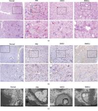
          Diabetic nephropathy (DN) is a major cause of chronic kidney disease. It has been proven that mesenchymal stem cells (MSCs) have therapeutic effects on kidney disease. Stem cells from human exfoliated deciduous teeth (SHED) are MSCs that are derived from dental pulps in exfoliated deciduous teeth from young patients and therefore have a high proliferation rate and an easy access. Hence, we aimed to explore the effect of SHED on DN in Goto-Kakizaki (GK) rats. SHED were administered via the tail vein. Blood glucose, serum triglycerides and cholesterol, body weight, and urinary albumin were measured before and after administration. At 8 weeks after administration, real-time PCR, immunohistochemistry (IHC), and electron microscopy were employed to examine pathological changes in glomerular and tubulointerstitial tissue. Kidney weight and serum IL-1, IL-10, TNF- α, TGF- β, and HGF levels were measured. SHED engraftment in the kidneys was detected by transfecting green fluorescence protein (GFP). Type II epithelial-mesenchymal transition (EMT) in the tubule-interstitial and arteriolar regions has been reported to be an important pathological characteristic of DN. This study is the first to apply a transwell system for coculture to explore the effects of MSCs on the EMT of human proximal tubular epithelial (HK-2) cells. The effects of SHED on advanced glycation end product- (AGE-) activated EMT in HK-2 cells were explored by real-time PCR and western blot. At 8 weeks after administration, renal injury, including hyperglycemia, hyperlipidemia, increased urinary albumin excretion, ECM accumulation, and a fractional mesangial area, was dramatically attenuated. The serum levels of IL-1, TNF- α, and TGF- β were significantly downregulated, whereas the serum levels of IL-10 and HGF were upregulated by SHED. GFP expression confirmed the engraftment of SHED in diabetic kidneys. In addition, cocultured SHED inhibited AGE-induced EMT in HK-2 cells. In conclusion, SHED offer a novel potential effective therapeutic approach for ameliorating DN.

          Related collections

          Most cited references40

          • Record: found
          • Abstract: found
          • Article: not found

          Pathophysiology and treatment of type 2 diabetes: perspectives on the past, present, and future.

          Glucose metabolism is normally regulated by a feedback loop including islet β cells and insulin-sensitive tissues, in which tissue sensitivity to insulin affects magnitude of β-cell response. If insulin resistance is present, β cells maintain normal glucose tolerance by increasing insulin output. Only when β cells cannot release sufficient insulin in the presence of insulin resistance do glucose concentrations rise. Although β-cell dysfunction has a clear genetic component, environmental changes play an essential part. Modern research approaches have helped to establish the important role that hexoses, aminoacids, and fatty acids have in insulin resistance and β-cell dysfunction, and the potential role of changes in the microbiome. Several new approaches for treatment have been developed, but more effective therapies to slow progressive loss of β-cell function are needed. Recent findings from clinical trials provide important information about methods to prevent and treat type 2 diabetes and some of the adverse effects of these interventions. However, additional long-term studies of drugs and bariatric surgery are needed to identify new ways to prevent and treat type 2 diabetes and thereby reduce the harmful effects of this disease. Copyright © 2014 Elsevier Ltd. All rights reserved.
            Bookmark
            • Record: found
            • Abstract: found
            • Article: not found

            Mechanisms of mesenchymal stromal cell immunomodulation.

            Multipotent mesenchymal stromal cells (MSCs) have generated considerable interest in the fields of regenerative medicine, cell therapy and immune modulation. Over the past 5 years, the initial observations that MSCs could enhance regeneration and modulate immune responses have been significantly advanced and we now have a clearer picture of the effects that MSCs have on the immune system particularly in the context of inflammatory-mediated disorders. A number of mechanisms of action have been reported in MSC immunomodulation, which encompass the secretion of soluble factors, induction of anergy, apoptosis, regulatory T cells and tolerogenic dendritic cells. It is clear that MSCs modulate both innate and adaptive responses and evidence is now emerging that the local microenvironment is key in the activation or licensing of MSCs to become immunosuppressive. More recently, studies have suggested that MSCs have the capacity to sense their environment and have a role in pathogen clearance in conjunction with the resolution of insult or injury. This review focuses on the mechanisms of MSC immunomodulation discussing the multistep process of MSC localisation at sites of inflammation, the cross talk between MSCs and the local microenvironment as well as the subsequent mechanisms of action used to resolve inflammation.
              Bookmark
              • Record: found
              • Abstract: found
              • Article: not found

              Multipotent stromal cells from human marrow home to and promote repair of pancreatic islets and renal glomeruli in diabetic NOD/scid mice.

              We tested the hypothesis that multipotent stromal cells from human bone marrow (hMSCs) can provide a potential therapy for human diabetes mellitus. Severe but nonlethal hyperglycemia was produced in NOD/scid mice with daily low doses of streptozotocin on days 1-4, and hMSCs were delivered via intracardiac infusion on days 10 and 17. The hMSCs lowered blood glucose levels in the diabetic mice on day 32 relative to untreated controls (18.34 mM +/- 1.12 SE vs. 27.78 mM +/- 2.45 SE, P = 0.0019). ELISAs demonstrated that blood levels of mouse insulin were higher in the hMSC-treated as compared with untreated diabetic mice, but human insulin was not detected. PCR assays detected human Alu sequences in DNA in pancreas and kidney on day 17 or 32 but not in other tissues, except heart, into which the cells were infused. In the hMSC-treated diabetic mice, there was an increase in pancreatic islets and beta cells producing mouse insulin. Rare islets contained human cells that colabeled for human insulin or PDX-1. Most of the beta cells in the islets were mouse cells that expressed mouse insulin. In kidneys of hMSC-treated diabetic mice, human cells were found in the glomeruli. There was a decrease in mesangial thickening and a decrease in macrophage infiltration. A few of the human cells appeared to differentiate into glomerular endothelial cells. Therefore, the results raised the possibility that hMSCs may be useful in enhancing insulin secretion and perhaps improving the renal lesions that develop in patients with diabetes mellitus.
                Bookmark

                Author and article information

                Contributors
                Journal
                Stem Cells Int
                Stem Cells Int
                SCI
                Stem Cells International
                Hindawi
                1687-966X
                1687-9678
                2019
                1 December 2019
                : 2019
                : 2751475
                Affiliations
                1Department of Pediatric Dentistry, Peking University School and Hospital of Stomatology, 22 Zhongguancun Avenue South, Haidian District, Beijing 100081, China
                2Department of Oral Emergency Department, Peking University School and Hospital of Stomatology, 22 Zhongguancun Avenue South, Haidian District, Beijing 100081, China
                3Department of Stomatology, Shenzhen Children's Hospital, No. 7019, Yitian Road, Shenzhen 518026, China
                Author notes

                Academic Editor: Mustapha Najimi

                Author information
                https://orcid.org/0000-0001-9770-2460
                https://orcid.org/0000-0002-2395-6254
                https://orcid.org/0000-0002-0106-7659
                Article
                10.1155/2019/2751475
                6913167
                f7c1e7c8-2978-49d5-8390-5211b72c2ab3
                Copyright © 2019 Nanquan Rao et al.

                This is an open access article distributed under the Creative Commons Attribution License, which permits unrestricted use, distribution, and reproduction in any medium, provided the original work is properly cited.

                History
                : 30 November 2018
                : 6 May 2019
                : 20 September 2019
                Funding
                Funded by: Peking University
                Award ID: 94000-6672-H78
                Funded by: National Natural Science Foundation of China
                Award ID: 303076129
                Categories
                Research Article

                Molecular medicine
                Molecular medicine

                Comments

                Comment on this article