33
views
0
recommends
+1 Recommend
0 collections
    0
    shares
      • Record: found
      • Abstract: found
      • Article: found
      Is Open Access

      The Extract of Aster Koraiensis Prevents Retinal Pericyte Apoptosis in Diabetic Rats and Its Active Compound, Chlorogenic Acid Inhibits AGE Formation and AGE/RAGE Interaction

      research-article

      Read this article at

      Bookmark
          There is no author summary for this article yet. Authors can add summaries to their articles on ScienceOpen to make them more accessible to a non-specialist audience.

          Abstract

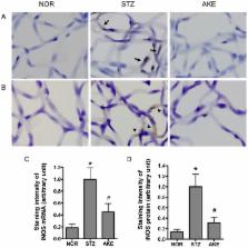
          Retinal capillary cell loss is a hallmark of early diabetic retinal changes. Advanced glycation end products (AGEs) are believed to contribute to retinal microvascular cell loss in diabetic retinopathy. In this study, the protective effects of Aster koraiensis extract (AKE) against damage to retinal vascular cells were investigated in streptozotocin (STZ)-induced diabetic rats. To examine this issue further, AGE accumulation, nuclear factor-kappaB (NF-κB) and inducible nitric oxide synthase (iNOS) were investigated using retinal trypsin digests from streptozotocin-induced diabetic rats. In the diabetic rats, TUNEL (Terminal deoxynucleotidyl transferase mediated dUTP Nick End Labeling)-positive retinal microvascular cells were markedly increased. Immunohistochemical studies revealed that AGEs were accumulated within the retinal microvascular cells, and this accumulation paralleled the activation of NF-κB and the expression of iNOS in the diabetic rats. However, AKE prevented retinal microvascular cell apoptosis through the inhibition of AGE accumulation and NF-κB activation. Moreover, to determine the active compounds of AKE, two major compounds, chlorogenic acid and 3,5-di- O-caffeoylquinic acid, were tested in an in vitro assay. Among these compounds, chlorogenic acid significantly reduced AGE formation as well as AGE/RAGE (receptor for AGEs) binding activity. These results suggest that AKE, particularly chlorogenic acid, is useful in inhibiting AGE accumulation in retinal vessels and exerts a preventive effect against the injuries of diabetic retinal vascular cells.

          Related collections

          Most cited references58

          • Record: found
          • Abstract: not found
          • Article: not found

          Nitric oxide synthases: roles, tolls, and controls.

            Bookmark
            • Record: found
            • Abstract: found
            • Article: not found

            Formation of glyoxal, methylglyoxal and 3-deoxyglucosone in the glycation of proteins by glucose.

            The glycation of proteins by glucose has been linked to the development of diabetic complications and other diseases. Early glycation is thought to involve the reaction of glucose with N-terminal and lysyl side chain amino groups to form Schiff's base and fructosamine adducts. The formation of the alpha-oxoaldehydes, glyoxal, methylglyoxal and 3-deoxyglucosone, in early glycation was investigated. Glucose (50 mM) degraded slowly at pH 7.4 and 37 degrees C to form glyoxal, methylglyoxal and 3-deoxyglucosone throughout a 3-week incubation period. Addition of t-BOC-lysine and human serum albumin increased the rate of formation of alpha-oxoaldehydes - except glyoxal and methylglyoxal concentrations were low with albumin, as expected from the high reactivity of glyoxal and methylglyoxal with arginine residues. The degradation of fructosyl-lysine also formed glyoxal, methylglyoxal and 3-deoxyglucosone. alpha-Oxoaldehyde formation was dependent on the concentration of phosphate buffer and availability of trace metal ions. This suggests that alpha-oxoaldehydes were formed in early glycation from the degradation of glucose and Schiff's base adduct. Since alpha-oxoaldehydes are important precursors of advanced glycation adducts, these adducts may be formed from early and advanced glycation processes. Short periods of hyperglycaemia, as occur in impaired glucose tolerance, may be sufficient to increase the concentrations of alpha-oxoaldehydes in vivo.
              Bookmark
              • Record: found
              • Abstract: found
              • Article: not found

              Role of transcription factor NF-kappa B/Rel in induction of nitric oxide synthase.

              The promoter of the murine gene encoding inducible nitric oxide synthase (iNOS) contains an NF-kappa B site beginning 55 base pairs upstream of the TATA box, designated NF-kappa Bd. Reporter constructs containing truncated promoter regions, when transfected into macrophages, revealed that NF-kappa Bd is necessary to confer inducibility by bacterial lipopolysaccharide (LPS). Oligonucleotide probes containing NF-kappa Bd plus the downstream 9 or 47 base pairs bound proteins that rapidly appeared in the nuclei of LPS-treated macrophages. The nuclear proteins bound to both probes in an NF-kappa Bd-dependent manner, but binding was resistant to cycloheximide only for the shorter probe. The proteins binding both probes reacted with antibodies against p50 and c-rel but not RelB; those binding the shorter probe also reacted with anti-RelA (p65). Pyrrolidine dithiocarbamate, which acts as a specific inhibitor of NF-kappa B, blocked both the activation of the NF-kappa Bd-binding proteins and the production of NO in LPS-treated macrophages. Thus, activation of NF-kappa B/Rel is critical in the induction of iNOS by LPS. However, additional, newly synthesized proteins contribute to the NF-kappa Bd-dependent transcription factor complex on the iNOS promoter in LPS-treated mouse macrophages.
                Bookmark

                Author and article information

                Journal
                Nutrients
                Nutrients
                nutrients
                Nutrients
                MDPI
                2072-6643
                21 September 2016
                September 2016
                : 8
                : 9
                : 585
                Affiliations
                Korean Medicine Convergence Research Division, Korea Institute of Oriental Medicine, Daejeon 34054, Korea; dvmhyun@ 123456kiom.re.kr (J.K.); jopd7414@ 123456kiom.re.kr (K.J.); knifer48@ 123456kiom.re.kr (I.-S.L.)
                Author notes
                [* ]Correspondence: chskim@ 123456kiom.re.kr (C.-S.K.); jskim@ 123456kiom.re.kr (J.S.K.); Tel.: +82-42-868-9473 (C.-S.K.); +82-42-868-9465 (J.S.K.); Fax: +82-42-868-9471 (C.-S.K. & J.S.K.)
                Article
                nutrients-08-00585
                10.3390/nu8090585
                5037569
                27657123
                f9336ac2-cb1b-493f-ab18-2d1ab9b5fa3c
                © 2016 by the authors; licensee MDPI, Basel, Switzerland.

                This article is an open access article distributed under the terms and conditions of the Creative Commons Attribution (CC-BY) license ( http://creativecommons.org/licenses/by/4.0/).

                History
                : 30 July 2016
                : 13 September 2016
                Categories
                Article

                Nutrition & Dietetics
                advanced glycation end products,diabetic retinopathy,aster koraiensis

                Comments

                Comment on this article