6
views
0
recommends
+1 Recommend
0 collections
    0
    shares
      • Record: found
      • Abstract: found
      • Article: found
      Is Open Access

      Targeting pyroptosis as a preventive and therapeutic approach for stroke

      review-article

      Read this article at

      Bookmark
          There is no author summary for this article yet. Authors can add summaries to their articles on ScienceOpen to make them more accessible to a non-specialist audience.

          Abstract

          Stroke has caused tremendous social stress worldwide, yet despite decades of research and development of new stroke drugs, most have failed and rt-PA (Recombinant tissue plasminogen activator) is still the accepted treatment for ischemic stroke. the complexity of the stroke mechanism has led to unsatisfactory efficacy of most drugs in clinical trials, indicating that there are still many gaps in our understanding of stroke. Pyroptosis is a programmed cell death (PCD) with inflammatory properties and are thought to be closely associated with stroke. Pyroptosis is regulated by the GSDMD of the gasdermin family, which when cleaved by Caspase-1/Caspase-11 into N-GSDMD with pore-forming activity can bind to the plasma membrane to form small 10–20 nm pores, which would allow the release of inflammatory factors IL-18 and IL-1β before cell rupture, greatly exacerbating the inflammatory response. The pyroptosis occurs mainly in the border zone of cerebral infarction, and glial cells, neuronal cells and brain microvascular endothelial cells (BMECs) all undergo pyroptosis after stroke, which largely exacerbates the breakdown of the blood-brain barrier (BBB) and thus aggravates brain injury. Therefore, pyroptosis may be a good direction for the treatment of stroke. In this review, we focus on the latest mechanisms of action of pyroptosis and the process by which pyroptosis regulates stroke development. We also suggest potential therapeutic stroke drugs that target the pyroptosis pathway, providing additional therapeutic strategies for the clinical management of stroke.

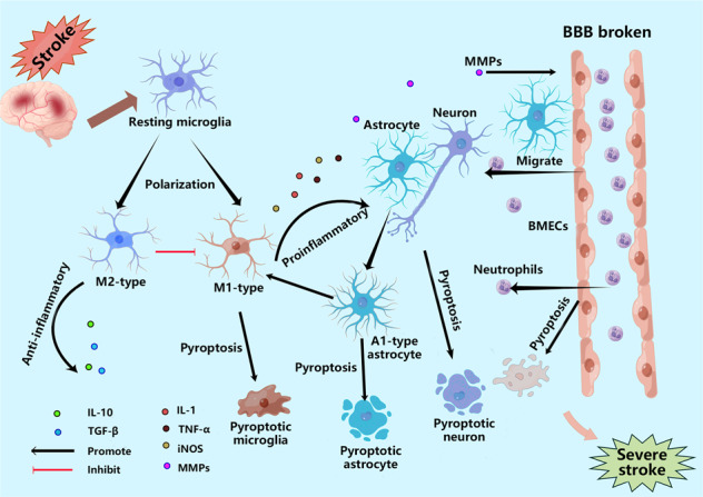
          The role of pyroptosis after stroke. After stroke, microglia first rush to the damaged area and polarize into M1 and M2 types. Under the influence of various stimuli, microglia undergo pyroptosis, release pro-inflammatory factors, and are converted to the M1 type; astrocytes and neuronal cells also undergo pyroptosis under the stimulation of various pro-inflammatory factors, leading to astrocyte death due to increased osmotic pressure in the membrane, resulting in water absorption and swelling until rupture. BMECs, the main structural component of the BBB, also undergo pyroptosis when stimulated by pro-inflammatory factors released from microglia and astrocytes, leading to the destruction of the structural integrity of the BBB, ultimately causing more severe brain damage. In addition, GSDMD in neutrophils mainly mediate the release of NETs rather than pyroptosis, which also aggravates brain injury. IL-10=interleukin-10; TGF-β = transforming growth factor-β; IL-18=interleukin-18; IL-1β = interleukin-1β; TNF-α = tumor necrosis factor-α; iNOS=induced nitrogen monoxide synthase; MMPs=Matrix metalloproteinases; GSDMD = gasdermin D; BMECs=brain microvascular endothelial cells; BBB = blood-brain barrier.

          Related collections

          Most cited references129

          • Record: found
          • Abstract: found
          • Article: not found

          Caspase-11 cleaves gasdermin D for non-canonical inflammasome signalling.

          Intracellular lipopolysaccharide from Gram-negative bacteria including Escherichia coli, Salmonella typhimurium, Shigella flexneri, and Burkholderia thailandensis activates mouse caspase-11, causing pyroptotic cell death, interleukin-1β processing, and lethal septic shock. How caspase-11 executes these downstream signalling events is largely unknown. Here we show that gasdermin D is essential for caspase-11-dependent pyroptosis and interleukin-1β maturation. A forward genetic screen with ethyl-N-nitrosourea-mutagenized mice links Gsdmd to the intracellular lipopolysaccharide response. Macrophages from Gsdmd(-/-) mice generated by gene targeting also exhibit defective pyroptosis and interleukin-1β secretion induced by cytoplasmic lipopolysaccharide or Gram-negative bacteria. In addition, Gsdmd(-/-) mice are protected from a lethal dose of lipopolysaccharide. Mechanistically, caspase-11 cleaves gasdermin D, and the resulting amino-terminal fragment promotes both pyroptosis and NLRP3-dependent activation of caspase-1 in a cell-intrinsic manner. Our data identify gasdermin D as a critical target of caspase-11 and a key mediator of the host response against Gram-negative bacteria.
            Bookmark
            • Record: found
            • Abstract: found
            • Article: not found

            Inflammasome-activated gasdermin D causes pyroptosis by forming membrane pores.

            Inflammatory caspases (caspases 1, 4, 5 and 11) are activated in response to microbial infection and danger signals. When activated, they cleave mouse and human gasdermin D (GSDMD) after Asp276 and Asp275, respectively, to generate an N-terminal cleavage product (GSDMD-NT) that triggers inflammatory death (pyroptosis) and release of inflammatory cytokines such as interleukin-1β. Cleavage removes the C-terminal fragment (GSDMD-CT), which is thought to fold back on GSDMD-NT to inhibit its activation. However, how GSDMD-NT causes cell death is unknown. Here we show that GSDMD-NT oligomerizes in membranes to form pores that are visible by electron microscopy. GSDMD-NT binds to phosphatidylinositol phosphates and phosphatidylserine (restricted to the cell membrane inner leaflet) and cardiolipin (present in the inner and outer leaflets of bacterial membranes). Mutation of four evolutionarily conserved basic residues blocks GSDMD-NT oligomerization, membrane binding, pore formation and pyroptosis. Because of its lipid-binding preferences, GSDMD-NT kills from within the cell, but does not harm neighbouring mammalian cells when it is released during pyroptosis. GSDMD-NT also kills cell-free bacteria in vitro and may have a direct bactericidal effect within the cytosol of host cells, but the importance of direct bacterial killing in controlling in vivo infection remains to be determined.
              Bookmark
              • Record: found
              • Abstract: found
              • Article: not found

              Microglia Function in the Central Nervous System During Health and Neurodegeneration.

              Microglia are resident cells of the brain that regulate brain development, maintenance of neuronal networks, and injury repair. Microglia serve as brain macrophages but are distinct from other tissue macrophages owing to their unique homeostatic phenotype and tight regulation by the central nervous system (CNS) microenvironment. They are responsible for the elimination of microbes, dead cells, redundant synapses, protein aggregates, and other particulate and soluble antigens that may endanger the CNS. Furthermore, as the primary source of proinflammatory cytokines, microglia are pivotal mediators of neuroinflammation and can induce or modulate a broad spectrum of cellular responses. Alterations in microglia functionality are implicated in brain development and aging, as well as in neurodegeneration. Recent observations about microglia ontogeny combined with extensive gene expression profiling and novel tools to study microglia biology have allowed us to characterize the spectrum of microglial phenotypes during development, homeostasis, and disease. In this article, we review recent advances in our understanding of the biology of microglia, their contribution to homeostasis, and their involvement in neurodegeneration. Moreover, we highlight the complexity of targeting microglia for therapeutic intervention in neurodegenerative diseases. Expected final online publication date for the Annual Review of Immunology Volume 35 is April 26, 2017. Please see http://www.annualreviews.org/page/journal/pubdates for revised estimates.
                Bookmark

                Author and article information

                Contributors
                278339420@qq.com
                chennh@imm.ac.cn
                Journal
                Cell Death Discov
                Cell Death Discov
                Cell Death Discovery
                Nature Publishing Group UK (London )
                2058-7716
                10 May 2023
                10 May 2023
                2023
                : 9
                : 155
                Affiliations
                [1 ]GRID grid.488482.a, ISNI 0000 0004 1765 5169, Hunan Engineering Technology Center of Standardization and Function of Chinese Herbal Decoction Pieces, College of Pharmacy, , Hunan University of Chinese Medicine, ; Changsha, P. R. China
                [2 ]Department of Pharmacy, Changsha Hospital for Matemal & Child Health Care, Changsha, P. R. China
                [3 ]GRID grid.412643.6, ISNI 0000 0004 1757 2902, Department of Pharmacy, , The First Hospital of Lanzhou University, ; Lanzhou, P. R. China
                [4 ]GRID grid.506261.6, ISNI 0000 0001 0706 7839, State Key Laboratory of Bioactive Substances and Functions of Natural Medicines, Institute of Materia Medica & Neuroscience Center, , Chinese Academy of Medical Sciences and Peking Union Medical College, ; Beijing, P. R. China
                [5 ]GRID grid.411615.6, ISNI 0000 0000 9938 1755, Key Laboratory of Cosmetic, China National Light Industry, , Beijing Technology and Business University, ; Beijing, P. R. China
                [6 ]Department of Nephrology, Xiangtan Central Hospital, Xiangtan, P. R. China
                Author information
                http://orcid.org/0000-0002-1647-2825
                Article
                1440
                10.1038/s41420-023-01440-y
                10172388
                37165005
                fa1f6761-d322-4e79-8f78-f28038da4fc6
                © The Author(s) 2023

                Open Access This article is licensed under a Creative Commons Attribution 4.0 International License, which permits use, sharing, adaptation, distribution and reproduction in any medium or format, as long as you give appropriate credit to the original author(s) and the source, provide a link to the Creative Commons license, and indicate if changes were made. The images or other third party material in this article are included in the article’s Creative Commons license, unless indicated otherwise in a credit line to the material. If material is not included in the article’s Creative Commons license and your intended use is not permitted by statutory regulation or exceeds the permitted use, you will need to obtain permission directly from the copyright holder. To view a copy of this license, visit http://creativecommons.org/licenses/by/4.0/.

                History
                : 2 March 2023
                : 18 April 2023
                : 20 April 2023
                Funding
                Funded by: The Outstanding Youth Project of the Hunan Education Department (Grant No. 21B0354), Changsha Natural Science Foundation (Grant No. kq2202269), the Special Scientific and Technological Project for Comprehensive Utilization of Ampelopsis grossedentata Resources of Hunan Qiankun Biotechnology Co., Ltd. (Grant No. 212010), the Key Project of Hunan University of Chinese Medicine School level Scientific Research Fund (Grant No. 2019xjjj001), The Hunan University of Chinese Medicine First-class Disciple Construction Project of Chinese Material Medica, The Key Discipline of Biological Engineering of Hunan University of Chinese medicine [2018] No.3.
                Funded by: Innovation project of Hunan University of Traditional Chinese Medicine (2022CX12).
                Funded by: Hunan Natural Science Foundation (Grant No. 2022JJ40456).
                Funded by: Hunan Natural Science Foundation (Grant No. 2022JJ40313), the Key Project of Hunan University of Chinese Medicine School level Scientific Research Fund (Grant No. 2021XJJJ028).
                Funded by: Changsha Natural Science Foundation (Grant No. kq2014091), Hunan Natural Science Foundation (Grant No. 2021JJ30512), Hunan University of Chinese Medicine discipline construction project (22JBZ052), The First-class Discipline Construction Project of Chemical Engineering and Technology of Hunan University of Traditional Chinese Medicine.
                Funded by: Changsha Natural Science Foundation (Grant No. kq2014085).
                Funded by: The Hunan University of Chinese Medicine First-class Disciple Construction Project of Chinese Material Medica.
                Categories
                Review Article
                Custom metadata
                © Cell Death Differentiation Association (ADMC) 2023

                cell death in the nervous system,stroke
                cell death in the nervous system, stroke

                Comments

                Comment on this article