0
views
0
recommends
+1 Recommend
0 collections
    0
    shares
      • Record: found
      • Abstract: found
      • Article: not found

      Tailored alginate/PCL-gelatin-β-TCP membrane for guided bone regeneration

      , , , ,
      Biomedical Materials
      IOP Publishing

      Read this article at

      ScienceOpenPublisher
      Bookmark
          There is no author summary for this article yet. Authors can add summaries to their articles on ScienceOpen to make them more accessible to a non-specialist audience.

          Abstract

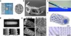
          Membranes prepared for guided bone regeneration (GBR) signify valued resources, inhibiting fibrosis and assisting bone regenration. However, existing membranes lack bone regenerative capacity or adequate degradation profile. An alginate-casted polycaprolactone-gelatin- β-tricalcium phosphate dual membrane was fabricated by electrospinning and casting processes to enhance new bone formation under a GBR process. Porous membranes were synthesized with suitable hydrophilicity, swelling, and degradation behavior to confirm the compatibility of the product in the body. Furthermore, osteoblast-type cell toxicity and cell adhesion results showed that the electrospun membrane offered compatible environment to cells while the alginate sheet was found capable enough to supress the cellular attachment, but was a non-toxic material. Post-implantation, the in-vivo outcomes of the dual-layered membrane, showed appreciable bone formation. Significantly, osteoid islands had fused in the membrane group by eight weeks. The infiltration of fibrous tissues was blocked by the alginate membrane, and the ingrowth of new bone was enhanced. Immunocytochemical analysis indicated that the dual membrane could direct more proteins which control mineralization and convene osteoconductive properties of tissue-engineered bone grafts.

          Related collections

          Most cited references45

          • Record: found
          • Abstract: found
          • Article: not found

          Engineering hydrogels as extracellular matrix mimics.

          Extracellular matrix (ECM) is a complex cellular environment consisting of proteins, proteoglycans, and other soluble molecules. ECM provides structural support to mammalian cells and a regulatory milieu with a variety of important cell functions, including assembling cells into various tissues and organs, regulating growth and cell-cell communication. Developing a tailored in vitro cell culture environment that mimics the intricate and organized nanoscale meshwork of native ECM is desirable. Recent studies have shown the potential of hydrogels to mimic native ECM. Such an engineered native-like ECM is more likely to provide cells with rational cues for diagnostic and therapeutic studies. The research for novel biomaterials has led to an extension of the scope and techniques used to fabricate biomimetic hydrogel scaffolds for tissue engineering and regenerative medicine applications. In this article, we detail the progress of the current state-of-the-art engineering methods to create cell-encapsulating hydrogel tissue constructs as well as their applications in in vitro models in biomedicine.
            Bookmark
            • Record: found
            • Abstract: found
            • Article: found
            Is Open Access

            Bioactive calcium phosphate materials and applications in bone regeneration

            Background Bone regeneration involves various complex biological processes. Many experiments have been performed using biomaterials in vivo and in vitro to promote and understand bone regeneration. Among the many biomaterials, calcium phosphates which exist in the natural bone have been conducted a number of studies because of its bone regenerative property. It can be directly contributed to bone regeneration process or assist in the use of other biomaterials. Therefore, it is widely used in many applications and has been continuously studied. Mainbody Calcium phosphate has been widely used in bone regeneration applications because it shows osteoconductive and in some cases osteoinductive features. The release of calcium and phosphorus ions regulates the activation of osteoblasts and osteoclasts to facilitate bone regeneration. The control of surface properties and porosity of calcium phosphate affects cell/protein adhesion and growth and regulates bone mineral formation. Properties affecting bioactivity vary depending on the types of calcium phosphates such as HAP, TCP and can be utilized in various applications because of differences in ion release, solubility, stability, and mechanical strength. In order to make use of these properties, different calcium phosphates have been used together or mixed with other materials to complement their disadvantages and to highlight their advantages. Calcium phosphate has been utilized to improve bone regeneration in ways such as increasing osteoconductivity for bone ingrowth, enhancing osteoinductivity for bone mineralization with ion release control, and encapsulating drugs or growth factors. Conclusion Calcium phosphate has been used for bone regeneration in various forms such as coating, cement and scaffold based on its unique bioactive properties and bone regeneration effectiveness. Additionally, several studies have been actively carried out to improve the efficacy of calcium phosphate in combination with various healing agents. By summarizing the properties of calcium phosphate and its research direction, we hope that calcium phosphate can contribute to the clinical treatment approach for bone defect and disease.
              Bookmark
              • Record: found
              • Abstract: found
              • Article: not found

              Biocompatibility and osteogenesis of biomimetic nano-hydroxyapatite/polyamide composite scaffolds for bone tissue engineering.

              In this study, we prepared nano-hydroxyapatite/polyamide (n-HA/PA) composite scaffolds utilizing thermally induced phase inversion processing technique. The macrostructure and morphology as well as mechanical strength of the scaffolds were characterized. Mesenchymal stem cells (MSCs) derived from bone marrow of neonatal rabbits were cultured, expanded and seeded on n-HA/PA scaffolds. The MSC/scaffold constructs were cultured for up to 7 days and the adhesion, proliferation and differentiation of MSCs into osteoblastic phenotype were determined using MTT assay, alkaline phosphatase (ALP) activity and collagen type I (COL I) immunohistochemical staining and scanning electronic microscopy (SEM). The results confirm that n-HA/PA scaffolds are biocompatible and have no negative effects on the MSCs in vitro. To investigate the in vivo biocompatibility and osteogenesis of the composite scaffolds, both pure n-HA/PA scaffolds and MSC/scaffold constructs were implanted in rabbit mandibles and studied histologically and microradiographically. The results show that n-HA/PA composite scaffolds exhibit good biocompatibility and extensive osteoconductivity with host bone. Moreover, the introduction of MSCs to the scaffolds dramatically enhanced the efficiency of new bone formation, especially at the initial stage after implantation. In long term (more than 12 weeks implantation), however, the pure scaffolds show as good biocompatibility and osteogenesis as the hybrid ones. All these results indicate that the scaffolds fulfill the basic requirements of bone tissue engineering scaffold, and have the potential to be applied in orthopedic, reconstructive and maxillofacial surgery.
                Bookmark

                Author and article information

                Contributors
                (View ORCID Profile)
                Journal
                Biomedical Materials
                Biomed. Mater.
                IOP Publishing
                1748-6041
                1748-605X
                May 17 2022
                July 01 2022
                May 17 2022
                July 01 2022
                : 17
                : 4
                : 045011
                Article
                10.1088/1748-605X/ac6bd8
                fb18b725-d255-4391-8ce4-b96e70766ae3
                © 2022

                https://iopscience.iop.org/page/copyright

                History

                Comments

                Comment on this article