18
views
0
recommends
+1 Recommend
0 collections
    0
    shares
      • Record: found
      • Abstract: found
      • Article: found
      Is Open Access

      The Correlation Between Preoperative Findings of High-Resolution Computed Tomography (HRCT) and Intraoperative Findings of Chronic Otitis Media (COM)

      research-article

      Read this article at

      Bookmark
          There is no author summary for this article yet. Authors can add summaries to their articles on ScienceOpen to make them more accessible to a non-specialist audience.

          Abstract

          Objectives:

          The aim of this study was to investigate the correlation between the preoperative findings of high-resolution computed tomography (HRCT) of temporal bone in chronic otitis media (COM) and the intraoperative findings.

          Methods:

          This retrospective study was conducted in the ORL-HNS Unit at Ohud Hospital, Medina, Saudi Arabia, during the period from January to September 2018. We included all patients with COM, and an informed consent was obtained from all participants. The HRCT images were studied in comparison with the intraoperative findings. The parameters of comparison were tympanic membrane, middle ear structures, and the status of cholesteatoma.

          Results:

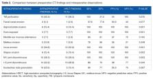
          A total of 39 patients were included in the analysis: 14 male and 25 female patients. The age range was 9 to 50 years. As a result of the comparison between HRCT findings and intraoperative observations, we found that incus erosion through computed tomography (CT) was the same as during surgical observation in 12 cases (30.8%). Malleus appeared eroded on CT in 1 case (2.6%); however, 5 cases were seen with that observation during operation (12.8%). Cholesteatoma was similarly seen in the CT scan and during surgery with a significant relation between intraoperative cholesteatoma extending and HRCT findings of the disease (95% confidence level, P-value = 0.001). The sensitivity, specificity, positive predictive value, and negative predictive value were 100% for detecting sigmoid plate erosion, dura exposure, incus erosion, stapes erosion, and malleus-incus joint discontinuity through preoperative CT.

          Conclusions:

          Intraoperative findings and HRCT have shown better results with good correlation of diagnostic value regarding the comparisons between recorded observations, especially in detecting sigmoidal plate erosion, dural exposure, incus and stapes erosion, and malleus-incus joint discontinuity. Preoperative CT scan is beneficial and contributory in the decision of indicating surgery to patients.

          Related collections

          Most cited references17

          • Record: found
          • Abstract: found
          • Article: not found

          Extracranial and intracranial complications of suppurative otitis media. Report of 102 cases.

          During the eight-year period, 1983-1990, 102 cases of intracranial (IC) and extracranial (EC) complications from 17 144 suppurative otitis media were reviewed. The prevalence of each complication was 0.24 and 0.45 per cent respectively. Facial paralysis, subperiosteal abscess and labyrinthitis were the common complications among the EC group, while meningitis and brain abscess were common in the IC group. Twenty five per cent of the EC group and 44 per cent of the IC group had more than two complications. The reliable warning signs and symptoms for IC complications were fever, headache, earache, vestibular symptoms, meningeal signs and impairment of consciousness. Proteus spp., Pseudomonas aeuruginosa and Staphylococcus spp. were the commonest organisms isolated from both groups. Cholesteatoma and granulation/polyp in the middle ear/mastoid were the major findings in both patient groups. Mortality rate in the IC group was 18.6 per cent. Morbidity rate in each group was 14.3 per cent (EC) and 27.9 per cent (IC) respectively. Epidemiological presentations, clinical features and the result of treatment are discussed.
            Bookmark
            • Record: found
            • Abstract: found
            • Article: not found

            Revision mastoidectomy.

            The first three priorities in surgery for chronic otitis media are (1) the elimination of progressive disease to produce a safe and dry ear, (2) modification of the anatomy of the tympanomastoid compartment to prevent recurrent disease, and (3) reconstruction of the hearing mechanism. The indications for revision following mastoidectomy for chronic otitis media thus involve failure to achieve any of these goals, including recurrent cholesteatoma, recurrent suppuration, recurrent perforation, or recurrent or residual conductive hearing loss. The focus of this article is the management of recurrent cholesteatoma or suppuration; that is, failure to achieve either of the first two priorities.
              Bookmark
              • Record: found
              • Abstract: found
              • Article: found
              Is Open Access

              Comparison of Preoperative Temporal Bone CT with Intraoperative Findings in Patients with Cholesteatoma

              Introduction: Cholesteatoma is traditionally diagnosed by otoscopic examination and treated by surgery. The necessity for imaging in an uncomplicated case is controversial. This study was planned to investigate the usefulness of a preoperative high-resolution computed tomography (HRCT) scan in depicting the status of middle ear structures in the presence of cholesteatoma and also to compare the correspondence between pre- and intraoperative CT findings in patients with cholesteatoma. Materials and Methods: This prospective descriptive study was performed from January 2009 to May 2011 in 36 patients with cholesteatoma who were referred to the Kashani and Al-Zahra Clinics of Otolaryngology. Preoperative high-resolution temporal bone CT scans (axial and coronal views) were carried out and compared with intraoperative findings. Results: Evaluation of 36 patients and their CT scans revealed excellent correlation for sigmoid plate erosion, widening of aditus, and erosion of scutum; good correlation for erosion of malleus and tegmen; moderate correlation for lateral canal fistula (LCF) and erosion of mastoid air cells; and poor correlation for facial nerve dehiscence (FND), incus, and stapes erosion. Conclusion: A preoperative CT scan may be helpful in relation to diagnosis and decision making for surgery in cases of cholesteatoma and ossicular erosion. The CT scan can accurately predict the extent of disease and is helpful for detection of lateral canal fistula, erosions of dural plate, and ossicular erosions. However it is not able to distinguish between cholesteatoma and mucosal disease, facial nerve dehiscency, incus, and stapes erosion.
                Bookmark

                Author and article information

                Journal
                Clin Med Insights Ear Nose Throat
                Clin Med Insights Ear Nose Throat
                ENT
                spent
                Clinical Medicine Insights. Ear, Nose and Throat
                SAGE Publications (Sage UK: London, England )
                1179-5506
                19 August 2019
                2019
                : 12
                : 1179550619870471
                Affiliations
                [1 ]Department of Otorhinolaryngology, Head and Neck surgery, Ohud Hospital, Madinah, Saudi Arabia
                [2 ]Department of Otology , Neurotology and Skull base surgery ,Ohud Hospital,Madinah,Saudi Arabia
                Author notes
                [*]Mariam Aljehani, Department of Otorhinolaryngology, Ohud Hospital, Salam District, Madinah 42354, Saudi Arabia. Email: marim_2012@ 123456hotmail.com
                Author information
                https://orcid.org/0000-0002-5826-2586
                Article
                10.1177_1179550619870471
                10.1177/1179550619870471
                6700852
                fc2730ff-2712-4f6c-8c6d-a50923368c9e
                © The Author(s) 2019

                This article is distributed under the terms of the Creative Commons Attribution-NonCommercial 4.0 License ( http://www.creativecommons.org/licenses/by-nc/4.0/) which permits non-commercial use, reproduction and distribution of the work without further permission provided the original work is attributed as specified on the SAGE and Open Access pages ( https://us.sagepub.com/en-us/nam/open-access-at-sage).

                History
                : 12 June 2019
                : 27 July 2019
                Categories
                Original Research
                Custom metadata
                January-December 2019

                temporal bone,chronic otitis media,computed tomography

                Comments

                Comment on this article