27
views
0
recommends
+1 Recommend
0 collections
    0
    shares
      • Record: found
      • Abstract: found
      • Article: found
      Is Open Access

      RLN3/RXFP3 Signaling in the PVN Inhibits Magnocellular Neurons via M-like Current Activation and Contributes to Binge Eating Behavior

      research-article

      Read this article at

      Bookmark
          There is no author summary for this article yet. Authors can add summaries to their articles on ScienceOpen to make them more accessible to a non-specialist audience.

          Abstract

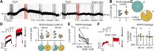
          Binge-eating disorder is the most common eating disorder. Various neuropeptides play important roles in the regulation of feeding behavior, including relaxin-3 (RLN3), which stimulates food intake in rats through the activation of the relaxin-family peptide-3 receptor (RXFP3). Here we demonstrate that a likely mechanism underlying the orexigenic action of RLN3 is RXFP3-mediated inhibition of oxytocin- and arginine-vasopressin-synthesizing paraventricular nucleus (PVN) magnocellular neurosecretory cells. Moreover, we reveal that, in male and female rats, this action depends on M-like potassium conductance. Notably, higher intra- and peri-PVN RLN3 fiber densities were observed in females, which may constitute an anatomic substrate for observed sex differences in binge-eating disorder. Finally, in a model of binge-eating in female rats, RXFP3 blockade within the PVN prevented binge-eating behavior. These data demonstrate a direct RLN3/RXFP3 action in the PVN of male and female rats, identify the associated ionic mechanisms, and reveal that hypothalamic RLN3/RXFP3 signaling regulates binge-eating behavior.

          SIGNIFICANCE STATEMENT Binge-eating disorder is the most common eating disorder worldwide, affecting women twice as frequently as men. Various neuropeptides play important roles in the regulation of feeding behavior, including relaxin-3, which acts via the relaxin-family peptide-3 receptor (RXFP3). Using a model of binge-eating, we demonstrated that relaxin-3/RXFP3 signaling in the hypothalamic paraventricular nucleus (PVN) is necessary for the expression of binge-eating behavior in female rats. Moreover, we elucidated the neuronal mechanism of RLN3/RXFP3 signaling in PVN in male and female rats and characterized sex differences in the RLN3 innervation of the PVN. These findings increase our understanding of the brain circuits and neurotransmitters involved in binge-eating disorder pathology and identify RXFP3 as a therapeutic target for binge-like eating disorders.

          Related collections

          Most cited references62

          • Record: found
          • Abstract: found
          • Article: not found

          Pathways modulating neural KCNQ/M (Kv7) potassium channels.

          K(+) channels play a crucial role in regulating the excitability of neurons. Many K(+) channels are, in turn, regulated by neurotransmitters. One of the first neurotransmitter-regulated channels to be identified, some 25 years ago, was the M channel. This was categorized as such because its activity was inhibited through stimulation of muscarinic acetylcholine receptors. M channels are now known to be composed of subunits of the Kv7 (KCNQ) K(+) channel family. However, until recently, the link between the receptors and the channels has remained elusive. Here, we summarize recent developments that have begun to clarify this link and discuss their implications for physiology and medicine.
            Bookmark
            • Record: found
            • Abstract: found
            • Article: not found

            Vasopressin and oxytocin receptor systems in the brain: Sex differences and sex-specific regulation of social behavior.

            The neuropeptides vasopressin (VP) and oxytocin (OT) and their receptors in the brain are involved in the regulation of various social behaviors and have emerged as drug targets for the treatment of social dysfunction in several sex-biased neuropsychiatric disorders. Sex differences in the VP and OT systems may therefore be implicated in sex-specific regulation of healthy as well as impaired social behaviors. We begin this review by highlighting the sex differences, or lack of sex differences, in VP and OT synthesis in the brain. We then discuss the evidence showing the presence or absence of sex differences in VP and OT receptors in rodents and humans, as well as showing new data of sexually dimorphic V1a receptor binding in the rat brain. Importantly, we find that there is lack of comprehensive analysis of sex differences in these systems in common laboratory species, and we find that, when sex differences are present, they are highly brain region- and species-specific. Interestingly, VP system parameters (VP and V1aR) are typically higher in males, while sex differences in the OT system are not always in the same direction, often showing higher OT expression in females, but higher OT receptor expression in males. Furthermore, VP and OT receptor systems show distinct and largely non-overlapping expression in the rodent brain, which may cause these receptors to have either complementary or opposing functional roles in the sex-specific regulation of social behavior. Though still in need of further research, we close by discussing how manipulations of the VP and OT systems have given important insights into the involvement of these neuropeptide systems in the sex-specific regulation of social behavior in rodents and humans.
              Bookmark
              • Record: found
              • Abstract: found
              • Article: not found

              Relaxin family peptides and their receptors.

              There are seven relaxin family peptides that are all structurally related to insulin. Relaxin has many roles in female and male reproduction, as a neuropeptide in the central nervous system, as a vasodilator and cardiac stimulant in the cardiovascular system, and as an antifibrotic agent. Insulin-like peptide-3 (INSL3) has clearly defined specialist roles in male and female reproduction, relaxin-3 is primarily a neuropeptide involved in stress and metabolic control, and INSL5 is widely distributed particularly in the gastrointestinal tract. Although they are structurally related to insulin, the relaxin family peptides produce their physiological effects by activating a group of four G protein-coupled receptors (GPCRs), relaxin family peptide receptors 1-4 (RXFP1-4). Relaxin and INSL3 are the cognate ligands for RXFP1 and RXFP2, respectively, that are leucine-rich repeat containing GPCRs. RXFP1 activates a wide spectrum of signaling pathways to generate second messengers that include cAMP and nitric oxide, whereas RXFP2 activates a subset of these pathways. Relaxin-3 and INSL5 are the cognate ligands for RXFP3 and RXFP4 that are closely related to small peptide receptors that when activated inhibit cAMP production and activate MAP kinases. Although there are still many unanswered questions regarding the mode of action of relaxin family peptides, it is clear that they have important physiological roles that could be exploited for therapeutic benefit.
                Bookmark

                Author and article information

                Journal
                J Neurosci
                J. Neurosci
                jneuro
                jneurosci
                J. Neurosci
                The Journal of Neuroscience
                Society for Neuroscience
                0270-6474
                1529-2401
                8 July 2020
                8 July 2020
                : 40
                : 28
                : 5362-5375
                Affiliations
                [1] 1Department of Neurophysiology and Chronobiology, Institute of Zoology and Biomedical Research, Faculty of Biology, Jagiellonian University, Krakow, 30-387, Poland
                [2] 2Department of Physical Biochemistry, Faculty of Biochemistry, Biophysics and Biotechnology, Jagiellonian University, Krakow, 30-387, Poland
                [3] 3School of Pharmacy, Pharmacology Unit, University of Camerino, Camerino, 62032, Italy
                [4] 4Florey Institute of Neuroscience and Mental Health, and Florey Department of Neuroscience and Mental Health, University of Melbourne, Melbourne, 3010 Victoria, Australia
                Author notes
                Correspondence should be addressed to Anna Blasiak at anna.blasiak@ 123456uj.edu.pl

                Author contributions: A.K., C.C., G.H., A.L.G., and A.B. designed research; A.K., A.S., P.S., A.G., E.B., and M.V.M.D.B. performed research; A.K., A.S., P.S., A.G., E.B., and M.V.M.D.B. analyzed data; A.K. and A.B. wrote the first draft of the paper; A.K., C.C., G.H., A.L.G., and A.B. edited the paper; M.A.H. and A.L.G. contributed unpublished reagents/analytic tools.

                Author information
                https://orcid.org/0000-0002-3823-6196
                https://orcid.org/0000-0002-7380-7011
                https://orcid.org/0000-0002-7457-2314
                https://orcid.org/0000-0003-0045-9025
                https://orcid.org/0000-0001-5936-064X
                https://orcid.org/0000-0002-8044-1206
                https://orcid.org/0000-0002-6066-9692
                https://orcid.org/0000-0002-4017-2988
                Article
                JN-RM-2895-19
                10.1523/JNEUROSCI.2895-19.2020
                7343322
                32532885
                fcab9ab9-2c01-45a7-b682-0730fd44ba38
                Copyright © 2020 Kania et al.

                This is an open-access article distributed under the terms of the Creative Commons Attribution License Creative Commons Attribution 4.0 International, which permits unrestricted use, distribution and reproduction in any medium provided that the original work is properly attributed.

                History
                : 10 December 2019
                : 17 April 2020
                : 21 May 2020
                Funding
                Funded by: Ministry of Science and Higher Education Poland
                Award ID: 0020/DIA/2014/43
                Funded by: The National Science Centre Poland
                Award ID: DEC-2012/05/D/NZ4/02984
                Award ID: UMO-2016/21/B/NZ4/00204
                Award ID: UMO-2017/24/T/NZ4/00225
                Funded by: Institute of Zoology and Biomedical Research of the Jagiellonian University
                Award ID: DS/MND/WBiNoZ/IZ/20/2016 - K/DSC/003960
                Award ID: DS/MND/WBiNoZ/IZ/16/2017 - K/DSC/004650
                Award ID: DS/MND/WB/IZ/9/2018 - K/DSC/005535
                Funded by: http://doi.org/10.13039/501100000925Department of Health | National Health and Medical Research Council (NHMRC)
                Award ID: 1122170
                Award ID: 1067522
                Funded by: Sir Edward ‘Weary’ Dunlop Research Fundation Mental Health Grant
                Funded by: Italian Ministry of Education, University and Research
                Award ID: PRIN2015KP7T2Y
                Categories
                Research Articles
                Cellular/Molecular
                Custom metadata
                true
                cellular

                binge eating,m-like current,paraventricular nucleus of hypothalamus,relaxin-3,rxfp3

                Comments

                Comment on this article