26
views
0
recommends
+1 Recommend
0 collections
    0
    shares
      • Record: found
      • Abstract: found
      • Article: found
      Is Open Access

      Sclerosing angiomatoid nodular transformation (SANT) of the spleen: a case report with FDG-PET findings and literature review

      case-report

      Read this article at

      Bookmark
          There is no author summary for this article yet. Authors can add summaries to their articles on ScienceOpen to make them more accessible to a non-specialist audience.

          Abstract

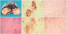
          We report the 18F-fluorodeoxyglucose (FDG) positron emission tomography (PET)/computed tomography (CT) findings of sclerosing angiomatoid nodular transformation (SANT) of the spleen. The patient was a 37-year-old woman with a splenic mass incidentally found on abdominal ultrasound. FDG-PET/CT showed weak FDG accumulation (maximum standardized uptake value = 3.65). An unenhanced CT scan showed a low density and well-circumscribed splenic tumor that demonstrated weak enhancement from the arterial to delayed phase. Although hemangioma or hamartoma of the spleen was preoperatively diagnosed, histopathological examination revealed SANT. Therefore, when a splenic tumor with weak contrast medium enhancement and low FDG accumulation is observed, SANT should be considered as a differential diagnosis. Although CT and magnetic resonance imaging features of SANT have been reported, there are few reports on FDG-PET/CT findings. We report the radiological features of SANT, including FDG-PET/CT, and review the literature on SANT.

          Related collections

          Most cited references13

          • Record: found
          • Abstract: found
          • Article: not found

          Sclerosing angiomatoid nodular transformation (SANT): report of 25 cases of a distinctive benign splenic lesion.

          Twenty-five cases of a morphologically distinctive vascular lesion of the spleen are described. The patients were 17 women and 8 men, ranging in age from 22 to 74 years (mean, 48.4 years; median, 56 years). The most common presentations were incidental finding of an asymptomatic splenic mass (13 patients), abdominal pain or discomfort (6 patients), and splenomegaly (4 patients). None of the patients had evidence of recurrent disease after splenectomy. The splenic lesion was solitary, measuring 3 to 17 cm, and sharply demarcated from the surrounding parenchyma. The cut surface revealed a mass of coalescing red-brown nodules embedded in a dense fibrous stroma. All cases showed a remarkably consistent multinodular appearance at low-power examination. The individual nodules had an angiomatoid appearance, in the sense that they were composed of slit-like, round or irregular-shaped vascular spaces lined by plump endothelial cells and interspersed by a population of spindly or ovoid cells. Some of the nodules (particularly the smaller ones) were surrounded by concentric rings of collagen fibers. Numerous red blood cells were present, as well as scattered inflammatory cells. Nuclear atypia was minimal, mitotic figures were extremely rare, and necrosis was consistently absent. The internodular stroma consisted of variably myxoid to dense fibrous tissue with scattered plump myofibroblasts, plasma cells, lymphocytes, and siderophages. Immunostaining revealed 3 distinct types of vessels in the angiomatoid nodules: CD34+/CD8-/CD31+ capillaries, CD34-/CD8+/CD31+ sinusoids, and CD34-/CD8-/CD31+ small veins, recapitulating the composition of the normal splenic red pulp. These features are therefore different from those of littoral cell angioma, conventional hemangioma, and hemangioendothelioma of the spleen. We interpret these angiomatoid nodules as altered red pulp tissue that had been entrapped by a nonneoplastic stromal proliferative process. The characteristic morphologic appearance, immunophenotype, and benign clinical course suggest that this is a distinctive nonneoplastic vascular lesion of the spleen that we propose to designate as sclerosing angiomatoid nodular transformation (SANT).
            Bookmark
            • Record: found
            • Abstract: found
            • Article: not found

            Percutaneous image-guided biopsy of the spleen: systematic review and meta-analysis of the complication rate and diagnostic accuracy.

            To use meta-analysis to determine the complication rate and diagnostic accuracy of image-guided percutaneous needle biopsy of the spleen. Several electronic databases were searched through July 2010 without language restrictions. Two reviewers independently selected studies that met the inclusion criteria for the diagnostic accuracy and complication rate arms of the study. Study data were independently extracted by the two reviewers. The primary 2 × 2 data were investigated with a random-effects meta-analysis of sensitivity and specificity. The complication rate data were investigated with a random-effects meta-analysis; sensitivity analysis of complication rate, excluding needles larger than 18 gauge, was performed. Four studies met the inclusion criteria for the diagnostic accuracy arm (639 patients), and nine met the inclusion criteria for the complication rate arm (741 patients). The meta-analysis showed a pooled sensitivity of 87.0% (95% confidence interval [CI]: 80.7%, 91.4%) and specificity of 96.4% (95% CI: 81.4%, 99.4%). The pooled major complication rate was 2.2% (95% CI: 0.8%, 5.6%). Sensitivity analysis with the removal of biopsies performed with needles larger than 18 gauge showed a major complication rate of 1.3% (95% CI: 0.6%, 2.5%). The most commonly encountered complications were hemorrhage followed by pain. Image-guided percutaneous biopsy of the spleen demonstrates high diagnostic accuracy and a major complication rate, for needles 18 gauge or smaller, that is similar to that reported for the liver and kidney. This technique should be considered a favorable alternative to splenectomy.
              Bookmark
              • Record: found
              • Abstract: found
              • Article: not found
              Is Open Access

              Sclerosing Angiomatoid Nodular Transformation (SANT) of the spleen: Case report and review of the literature.

              Sclerosing Angiomatoid Nodular Transformation of the spleen (SANT) is a rare benign vascular lesion of the spleen with extensive sclerosis and unknown etiology. We report a new case of SANT of the spleen found in a 53-year-old female following detection of a splenic mass on a routine computed tomography (CT). The patient underwent an uncomplicated laparoscopic splenectomy and the specimen was sent for histopathologic examination. A review of the 97 reported cases of SANT found in the literature was undertaken. There were 43 males and 54 females with a median age of 46 years (range: 11-82 years). SANT is classically considered to be a female predominant disease, however 44.3% of reported case were male and the gender predilection may soon be neutralized as more cases are reported. 65 of the 97 (67%) patients were in 30-60 year age group. The majority of lesions (n=50) were incidentally found on imaging, and for those patients presenting with symptoms, abdominal pain (n=18) was the predominant symptom. The diagnosis of SANT should be considered in any patient presenting with a splenic lesion that contains an angiomatoid or inflammatory component. As the differential diagnosis for SANT includes malignant pathologies, and currently no reliable diagnostic radiological feature has been identified to differentiate between these conditions, SANT will continue to be diagnosed on the basis of surgical histopathology. Copyright © 2012 Surgical Associates Ltd. Published by Elsevier Ltd. All rights reserved.
                Bookmark

                Author and article information

                Journal
                Acta Radiol Open
                Acta Radiol Open
                ARR
                sparr
                Acta Radiologica Open
                SAGE Publications (Sage UK: London, England )
                2058-4601
                10 August 2016
                August 2016
                : 5
                : 8
                : 2058460116649799
                Affiliations
                [1 ]Department of Diagnostic Imaging and Nuclear Medicine, Tokyo Women’s Medical University, Tokyo, Japan
                [2 ]Department of Surgery II, Tokyo Women’s Medical University, Tokyo, Japan
                [3 ]Department of Pathology, Tokyo Women’s Medical University, Tokyo, Japan
                Author notes
                [*]Reiko Nakajima, Department of Diagnostic Imaging and Nuclear Medicine, Tokyo Women’s Medical University, 8-1 Kawada-cho, Shinjuku-ku, Tokyo 162-8666, Japan. Email: nakajima.reiko@ 123456twmu.ac.jp
                Article
                10.1177_2058460116649799
                10.1177/2058460116649799
                4984319
                27570634
                fcb743a2-a260-4745-90f7-4906f8c4b2d0
                © The Foundation Acta Radiologica 2016

                This article is distributed under the terms of the Creative Commons Attribution-NonCommercial 3.0 License ( http://www.creativecommons.org/licenses/by-nc/3.0/) which permits non-commercial use, reproduction and distribution of the work without further permission provided the original work is attributed as specified on the SAGE and Open Access page ( https://us.sagepub.com/en-us/nam/open-access-at-sage).

                History
                : 25 March 2016
                : 22 April 2016
                Categories
                Case Report
                Custom metadata
                corrected-proof

                sclerosing angiomatoid nodular transformation (sant),spleen,18f-fluorodeoxyglucose (fdg) positron emission tomography (pet),computed tomography (ct)

                Comments

                Comment on this article