28
views
0
recommends
+1 Recommend
0 collections
    0
    shares
      • Record: found
      • Abstract: found
      • Article: found
      Is Open Access

      Protective role of microglial HO-1 blockade in aging: Implication of iron metabolism

      research-article
      a , b , a , b , a , b , c , c , a , b ,
      Redox Biology
      Elsevier
      Heme oxygenase-1, Microglia, Neuroinflammation, Iron metabolism, Ferroptosis, Aging, AD, Alzheimer's disease, ANOVA, analysis of variance, CD163, cluster of differentiation 163, CNS, central nervous system, CO, carbon monoxide, DFX, deferoxamine, DHE, dihydroethydium, DMT1, divalent metal transporter 1, EAE, experimental autoimmune encephalomyelitis, Fn, ferritin, FPN1, ferroportin 1, GPX4, glutathione peroxidase 4, Hb, hemoglobin, HCT, hematocrit, H2DCFDA, 2′,7′-dichlorodihydrofluorescein diacetate, HO-1, heme oxygenase 1, IL-1β, interleukin 1 beta, iNOS, inducible nitric oxide synthase, LPS, lipopolysaccharide, MCV, mean corpuscular volume, NDD, neurodegenerative diseases, NFT, neurofibrillary tangles, NLRP3, pyrin domain containing 3, NO, nitric oxide, NOR, novel object recognition test, NRF2, nuclear factor (erythroid-derived 2)-like 2, OS, oxidative stress, PD, Parkinson's disease, PI, propidium iodide, TFR1, transferrin receptor 1, TNF-α, tumor necrosis factor alpha, WBC, white cell counts, ZnPP, zinc protoporphyrin.

      Read this article at

      Bookmark
          There is no author summary for this article yet. Authors can add summaries to their articles on ScienceOpen to make them more accessible to a non-specialist audience.

          Abstract

          Heme oxygenase-1 (HO-1) is an inducible enzyme known for its anti-inflammatory, antioxidant and neuroprotective effects. However, increased expression of HO-1 during aging and age-related neurodegenerative diseases have been associated to neurotoxic ferric iron deposits. Being microglia responsible for the brain's innate immune response, the aim of this study was to understand the role of microglial HO-1 under inflammatory conditions in aged mice. For this purpose, aged wild type (WT) and LysMCreHmox1 △△ (HMOX1 M-KO) mice that lack HO-1 in microglial cells, were used. Aged WT mice showed higher basal expression levels of microglial HO-1 in the brain than adult mice. This increase was even higher when exposed to an inflammatory stimulus (LPS via i.p.) and was accompanied by alterations in different iron-related metabolism proteins, resulting in an increase of iron deposits, oxidative stress, ferroptosis and cognitive decline. Furthermore, microglia exhibited a primed phenotype and increased levels of inflammatory markers such as iNOS, p65, IL-1β, TNF-α, Caspase-1 and NLRP3. Interestingly, all these alterations were prevented in aged HMOX1 M-KO and WT mice treated with the HO-1 inhibitor ZnPPIX. In order to determine the effects of microglial HO-1-dependent iron overload, aged WT mice were treated with the iron chelator deferoxamine (DFX). DFX caused major improvements in iron, inflammatory and behavioral alterations found in aged mice exposed to LPS. In conclusion, this study highlights how microglial HO-1 overexpression contributes to neurotoxic iron accumulation providing deleterious effects in aged mice exposed to an inflammatory insult.
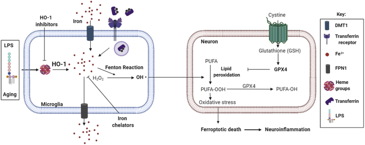
          Graphical abstract

          Highlights

          • Microglial HO-1 increases with aging and under an acute inflammatory stimulus.

          • LPS-dependent microglial HO-1 upregulation during aging leads to iron overload.

          • Microglial HO-1-dependent iron accumulation leads to ferroptosis.

          • HO-1-dependent iron alterations lead to neuroinflammation.

          • HO-1 inhibitors/iron chelators reduce iron accumulation and neuroinflammation.

          Related collections

          Most cited references82

          • Record: found
          • Abstract: found
          • Article: not found

          Ferroptosis: A Regulated Cell Death Nexus Linking Metabolism, Redox Biology, and Disease

          Ferroptosis is a form of regulated cell death characterized by the iron-dependent accumulation of lipid hydroperoxides to lethal levels. Emerging evidence suggests that ferroptosis represents an ancient vulnerability caused by the incorporation of polyunsaturated fatty acids into cellular membranes, and cells have developed complex systems that exploit and defend against this vulnerability in different contexts. The sensitivity to ferroptosis is tightly linked to numerous biological processes, including amino acid, iron, and polyunsaturated fatty acid metabolism, and the biosynthesis of glutathione, phospholipids, NADPH, and coenzyme Q10. Ferroptosis has been implicated in the pathological cell death associated with degenerative diseases (i.e., Alzheimer's, Huntington's, and Parkinson's diseases), carcinogenesis, stroke, intracerebral hemorrhage, traumatic brain injury, ischemia-reperfusion injury, and kidney degeneration in mammals and is also implicated in heat stress in plants. Ferroptosis may also have a tumor-suppressor function that could be harnessed for cancer therapy. This Primer reviews the mechanisms underlying ferroptosis, highlights connections to other areas of biology and medicine, and recommends tools and guidelines for studying this emerging form of regulated cell death.
            Bookmark
            • Record: found
            • Abstract: found
            • Article: not found

            Complement and microglia mediate early synapse loss in Alzheimer mouse models.

            Synapse loss in Alzheimer's disease (AD) correlates with cognitive decline. Involvement of microglia and complement in AD has been attributed to neuroinflammation, prominent late in disease. Here we show in mouse models that complement and microglia mediate synaptic loss early in AD. C1q, the initiating protein of the classical complement cascade, is increased and associated with synapses before overt plaque deposition. Inhibition of C1q, C3, or the microglial complement receptor CR3 reduces the number of phagocytic microglia, as well as the extent of early synapse loss. C1q is necessary for the toxic effects of soluble β-amyloid (Aβ) oligomers on synapses and hippocampal long-term potentiation. Finally, microglia in adult brains engulf synaptic material in a CR3-dependent process when exposed to soluble Aβ oligomers. Together, these findings suggest that the complement-dependent pathway and microglia that prune excess synapses in development are inappropriately activated and mediate synapse loss in AD.
              Bookmark
              • Record: found
              • Abstract: found
              • Article: not found

              Microglia and macrophages in brain homeostasis and disease

              Microglia and non-parenchymal macrophages in the brain are mononuclear phagocytes that are increasingly recognized to be essential players in the development, homeostasis and diseases of the central nervous system. With the availability of new genetic, molecular and pharmacological tools, considerable advances have been made towards our understanding of the embryonic origins, developmental programmes and functions of these cells. These exciting discoveries, some of which are still controversial, also raise many new questions, which makes brain macrophage biology a fast-growing field at the intersection of neuroscience and immunology. Here, we review the current knowledge of how and where brain macrophages are generated, with a focus on parenchymal microglia. We also discuss their normal functions during development and homeostasis, the disturbance of which may lead to various neurodegenerative and neuropsychiatric diseases.
                Bookmark

                Author and article information

                Contributors
                Journal
                Redox Biol
                Redox Biol
                Redox Biology
                Elsevier
                2213-2317
                06 November 2020
                January 2021
                06 November 2020
                : 38
                : 101789
                Affiliations
                [a ]Instituto Teófilo Hernando for Drug Discovery. Department of Pharmacology. School of Medicine. Universidad Autónoma Madrid. Madrid, Spain
                [b ]Instituto de Investigación Sanitario (IIS-IP), Hospital Universitario de La Princesa, Madrid, Spain
                [c ]Department of Anatomy, Histology and Neuroscience. School of Medicine. Universidad Autónoma de Madrid. Madrid, Spain
                Author notes
                []Corresponding author. Department of Pharmacology School of Medicine-UAM Calle Arzobispo Morcillo, 4 28029, Madrid, Spain. manuela.garcia@ 123456uam.es
                Article
                S2213-2317(20)30994-0 101789
                10.1016/j.redox.2020.101789
                7680814
                33212416
                fdd49829-66f8-4dc3-af60-a8c177afcea3
                © 2020 The Authors

                This is an open access article under the CC BY-NC-ND license (http://creativecommons.org/licenses/by-nc-nd/4.0/).

                History
                : 23 July 2020
                : 6 October 2020
                : 2 November 2020
                Categories
                Research Paper

                heme oxygenase-1,microglia,neuroinflammation,iron metabolism,ferroptosis,aging,ad, alzheimer's disease,anova, analysis of variance,cd163, cluster of differentiation 163,cns, central nervous system,co, carbon monoxide,dfx, deferoxamine,dhe, dihydroethydium,dmt1, divalent metal transporter 1,eae, experimental autoimmune encephalomyelitis,fn, ferritin,fpn1, ferroportin 1,gpx4, glutathione peroxidase 4,hb, hemoglobin,hct, hematocrit,h2dcfda, 2′,7′-dichlorodihydrofluorescein diacetate,ho-1, heme oxygenase 1,il-1β, interleukin 1 beta,inos, inducible nitric oxide synthase,lps, lipopolysaccharide,mcv, mean corpuscular volume,ndd, neurodegenerative diseases,nft, neurofibrillary tangles,nlrp3, pyrin domain containing 3,no, nitric oxide,nor, novel object recognition test,nrf2, nuclear factor (erythroid-derived 2)-like 2,os, oxidative stress,pd, parkinson's disease,pi, propidium iodide,tfr1, transferrin receptor 1,tnf-α, tumor necrosis factor alpha,wbc, white cell counts,znpp, zinc protoporphyrin.

                Comments

                Comment on this article