148
views
0
recommends
+1 Recommend
0 collections
    36
    shares
      • Record: found
      • Abstract: found
      • Article: found
      Is Open Access

      Re-operation of idiopathic full-thickness macular holes after initial surgery with internal limiting membrane peel

      research-article

      Read this article at

      Bookmark
          There is no author summary for this article yet. Authors can add summaries to their articles on ScienceOpen to make them more accessible to a non-specialist audience.

          Abstract

          Background/aims

          A retrospective consecutive case series to evaluate the efficacy of re-operation in patients with persistent or recurrent idiopathic full-thickness macular hole after initial surgery with internal limiting membrane peel (ILM).

          Methods

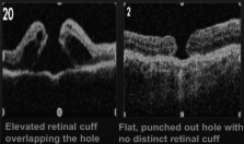
          491 patients underwent surgery for full-thickness macular hole from January 2004 to November 2007. Fifty-five patients either did not close or reopened during the follow-up period. Thirty patients with initial ILM peel underwent repeat surgery involving vitrectomy, enlargement of ILM rhexis and gas tamponade.

          Results

          Anatomical closure rate was 88.8% for primary surgery and 46.7% (14/30) for re-operation. There was a statistically significant improvement in overall best corrected visual acuity (BCVA) from re-operation baseline BCVA (p=0.02) within 1 year. For holes that did not close after the second surgery, visual acuity did not worsen.

          Conclusion

          Re-operation has a reduced success rate of anatomical closure. However, BCVA is statistically significantly improved from re-operation baseline, so even though we cannot return vision to pre-pathological baseline, re-operation can improve on this new baseline.

          Related collections

          Most cited references16

          • Record: found
          • Abstract: found
          • Article: not found

          Macular hole surgery with and without internal limiting membrane peeling.

          To compare results of surgery for idiopathic macular hole with and without internal limiting membrane (ILM) peeling in a series of consecutive patients over a 5-year period. A retrospective, nonrandomized, comparative trial with concurrent control group. Forty-four eyes with macular holes of less than or equal to 6 months duration without ILM peeling were compared to 116 eyes with ILM peeling and the same hole duration. A third group of 65 eyes with ILM peeling and duration greater than 6 months was also evaluated. All eyes underwent pars plana vitrectomy with or without ILM peeling, intravitreous gas, and positioning face down. No adjunctive therapies were used in any group. Comparing the closure and/or reopening rate, prognosis, visual acuity, and complications for macular holes with and without ILM peeling. All patients had postsurgical follow-up of 18 months or greater. Primary closure was significantly improved with ILM peeling with 116 of 116 eyes (100%) showing no reopenings versus 36 of 44 holes (82%) primarily closed, 9 of which (25%) reopened without ILM peeling (P: < 0.00001) in holes less than or equal to 6 months. The 27 eyes without ILM peeling that had successful surgery displayed a mean postoperative vision of 20/40, which is the same as the successful eyes with ILM peeling (P: = 0.6). The 52 stage II eyes with ILM peeling had a mean postoperative vision of 20/30, and 48 of the 52 eyes (92%) were 20/40 or better. Stage III eyes (greater than 400-microm holes) without ILM peeling had a poor prognosis, with 6 of the 25 eyes (24%) having initial surgery fail and an additional 4 of 25 eyes (16%) reopening. Without ILM peeling, holes less than 300 microm had only one reopen, whereas holes greater than or equal to 300 microm had 16 of the 17 (94%) primary failures and/or reopenings (P: < 0.001). All 12 holes that reopened and/or primarily failed were repaired with ILM peeling with excellent visual recovery. Macular holes with a duration greater than 6 months were treated with ILM peeling, and 63 of 65 holes (97%) were closed primarily and 65% had an increase in vision by two or more Snellen lines. ILM peeling significantly improves visual and anatomic success in all stages of recent and chronic macular holes and reopened and failed holes, while eliminating reopening for holes greater than 300 microm.
            Bookmark
            • Record: found
            • Abstract: found
            • Article: not found

            Vitreous surgery for macular holes.

            To surgically treat patients with macular holes, the authors previously reported both anatomic (re-attachment) and visual success (2 lines of improvement) in a series of 52 eyes. They now have operated on an additional 118 eyes using similar techniques, for a total of 170 eyes. After ophthalmologic examination and history, the authors operated on suitable patients. The surgical objectives included relief of all tangential traction and retinal tamponade with intraocular gas. All eyes were followed for at least 6 months postoperatively. In the total population of 170 eyes, anatomic success was achieved in 73% and vision improved at least two lines in 55%. Twenty-nine percent (49/170) of patients had a visual acuity of 20/40 or better at last examination. Patients with symptoms of less than 6 months' duration managed better than those with symptoms of longer duration (P = 0.3001). In the former group of 66 eyes, anatomic success was achieved in 80% (n = 53), whereas visual acuity improved at least two lines in 68% (n = 45) and at least four lines in 55% (n = 36). The authors suggest that macular hole surgery may provide meaningful improvement in visual acuity in most patients, especially in those whose symptoms are of less than 6 months' duration.
              Bookmark
              • Record: found
              • Abstract: found
              • Article: not found

              Focal macular ERGs in eyes after removal of macular ILM during macular hole surgery.

              The removal of the internal limiting membrane (ILM) for traction maculopathy has recently been advocated. However, it is generally believed that the ILM plays an important role in retinal function, because it is the basal lamina of the Müller cells that are involved in the generation of the electroretinogram (ERG) b-wave. To date, there has been no objective assessment of retinal function on removing the ILM. In this study, the changes of each component of the focal macular electroretinograms (FMERGs) were investigated in eyes before and after the ILM was removed in the macular area during surgery for idiopathic macular holes (IMHs). FMERGs were elicited by a 15 degrees stimulus centered on the fovea and monitored by an infrared fundus camera. FMERGs were recorded from 49 eyes of 48 patients with IMHs before and 6 weeks after anatomically successful macular hole surgery. Whether an eye had or did not have the ILM removed was randomly determined. The ILM was removed in 30 eyes (ILM-off group) and was not removed in 19 eyes (ILM-on group). Six months after surgery, the same examination was performed in 27 eyes of the ILM-off group and in 15 eyes of the ILM-on group. The amplitudes and implicit times of the a- and b-waves and the mean amplitudes and implicit times of the first three oscillatory potentials (OP1 to OP3) were compared before and after surgery within and between the groups. Visual acuity increased significantly after surgery in both groups. In the ILM-on group, the amplitude of the a- and b-waves and the OPs increased significantly 6 months after surgery (P: = 0.0093, P: = 0.0019, P: = 0.0024, respectively, paired t-test). In the ILM-off group, the a-wave amplitude and mean OP amplitudes were significantly larger 6 months after surgery (P: = 0.0077, P: = 0.0030, respectively, paired t-test). The b-wave amplitude, however, did not change significantly. The percentage increase in the b-wave amplitude 6 months after surgery was significantly higher in the ILM-on group (44.0%) than in the ILM-off group (15.0%; P: = 0.037, t-test). The removal of the ILM had no adverse effect on visual acuity. However, the selective delay of recovery of the FMERG b-wave 6 months after surgery suggests an alteration of retinal physiology in the macular region.
                Bookmark

                Author and article information

                Journal
                Br J Ophthalmol
                bjo
                bjophthalmol
                The British Journal of Ophthalmology
                BMJ Group (BMA House, Tavistock Square, London, WC1H 9JR )
                0007-1161
                1468-2079
                25 February 2011
                November 2011
                25 February 2011
                : 95
                : 11
                : 1564-1567
                Affiliations
                [1 ]Department of Family Medicine, University of Western Ontario, London, Ontario, Canada
                [2 ]Department of Surgery, St. Joseph Health Care, McMaster University, Hamilton, Ontario, Canada
                [3 ]Toronto Western Hospital, University of Toronto, Toronto, Ontario, Canada
                [4 ]The John and Liz Tory Eye Centre, Sunnybrook Health Sciences Centre, University of Toronto, Toronto, Ontario, Canada
                [5 ]Department of Ophthalmology and Vision Sciences, University of Toronto, Toronto, Ontario, Canada
                Author notes
                Correspondence to Dr Wai-Ching Lam, Department of Ophthalmology, East Wing, Room 6E 432, Toronto Western Hospital Toronto, Ontario, Canada M5T 2S8; waiching.lam@ 123456utoronto.ca
                Article
                bjophthalmol195826
                10.1136/bjo.2010.195826
                3199446
                21355018
                fdea2530-e32f-4e7c-891d-88f88b68ccb3
                © 2011, Published by the BMJ Publishing Group Limited. For permission to use (where not already granted under a licence) please go to http://group.bmj.com/group/rights-licensing/permissions.

                This is an open-access article distributed under the terms of the Creative Commons Attribution Non-commercial License, which permits use, distribution, and reproduction in any medium, provided the original work is properly cited, the use is non commercial and is otherwise in compliance with the license. See: http://creativecommons.org/licenses/by-nc/2.0/ and http://creativecommons.org/licenses/by-nc/2.0/legalcode.

                History
                : 30 January 2011
                Categories
                Clinical Science
                1506
                Original article

                Ophthalmology & Optometry
                indocyanine green,macula,internal limiting membrane peeling,vitrectomy,treatment surgery,re-operation,vision,vitreous,macular hole

                Comments

                Comment on this article