29
views
0
recommends
+1 Recommend
0 collections
    0
    shares
      • Record: found
      • Abstract: found
      • Article: found
      Is Open Access

      Bullous Systemic Lupus Erythematosus Associated with Esophagitis Dissecans Superficialis

      case-report

      Read this article at

      Bookmark
          There is no author summary for this article yet. Authors can add summaries to their articles on ScienceOpen to make them more accessible to a non-specialist audience.

          Abstract

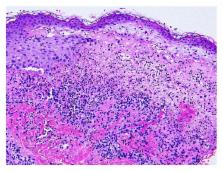
          Bullous systemic lupus erythematosus is one of the rare autoantibody mediated skin manifestation of systemic lupus erythematosus (SLE) demonstrating subepidermal blistering with neutrophilic infiltrate histologically. We present a case of a 40-year-old Hispanic female who presented with a several months' history of multiple blistering pruritic skin lesions involving the face and trunk, a photosensitive rash over the face and neck, swelling of the right neck lymph node, and joint pain involving her elbows and wrist. Her malady was diagnosed as bullous systemic lupus erythematosus based on the immunological workup and biopsy of her skin lesions. The patient also complained of odynophagia and endoscopy revealed esophagitis dissecans superficialis which is a rare endoscopic finding characterized by sloughing of the esophageal mucosa. The bullous disorders typically associated with esophagitis dissecans superficialis are pemphigus and rarely bullous pemphigoid. However, this is the first reported case of bullous systemic lupus erythematosus associated with esophagitis dissecans superficialis.

          Related collections

          Most cited references27

          • Record: found
          • Abstract: found
          • Article: not found

          Incidence and distribution of subepidermal autoimmune bullous skin diseases in three French regions. Bullous Diseases French Study Group.

          The incidence and distribution of autoimmune subepidermal bullous diseases were estimated from prospective data (including immunoelectron microscopy) obtained from 100 cases during a mean period of 35 months in three university dermatologic centers in Amiens, Limoges, and Tours, France, that correspond to a cumulative reference population of 3.55 x 10(6). Using data from these regions, we found a mean annual incidence of autoimmune subepidermal bullous diseases to be 10.4 per million people and, therefore, estimated the overall number of new cases of these disorders in France to be about 590 cases per year. According to clinical and immunoelectron microscopic criteria, a precise diagnosis was established in 94 cases, distributed as follows: bullous pemphigoid, 69 cases; cicatricial pemphigoid, 12 cases; linear IgA dermatosis, five cases; herpes gestationis, four cases; epidermolysis bullosa acquisita, two cases; and vesiculobullous systemic lupus erythematosus, two cases. Our prospective study is the first assessing the incidence and distribution of autoimmune subepidermal bullous disorders that systematically included immunoelectron microscopic data. Our estimated incidence of bullous pemphigoid (seven new cases per million people per year) is large enough to establish bullous pemphigoid as the major autoimmune subepidermal bullous disease for the purpose of therapeutic trials. On the contrary, all other disorders, particularly epidermolysis bullosa acquisita (estimated annual incidence, 0.17 to 0.26 per million people), were very rare and reflect the paucity of patients available for short-term clinical studies in France.
            Bookmark
            • Record: found
            • Abstract: found
            • Article: not found

            Identification of the skin basement-membrane autoantigen in epidermolysis bullosa acquisita.

            Epidermolysis bullosa acquisita is an acquired chronic blistering disease of the skin, in which separation of the skin occurs in the basement-membrane zone between the epidermis and the dermis. There is evidence that blistering is initiated by an immune process. Using serum samples from nine patients as a source of antibodies, we have identified a major protein of the basement membrane of human skin that serves as the antigen (or target) for autoantibodies in this disorder. This previously unrecognized protein, which consists of two components of 290,000 and 145,000 daltons, is distinct from other known components of the basement membrane. These studies provide evidence that epidermolysis bullosa acquisita is a specific disease that is different from other primary bullous diseases, such as bullous pemphigoid and pemphigus vulgaris, and suggest that the basement-membrane component that has been identified may have a role in normal epidermal-dermal adherence.
              Bookmark
              • Record: found
              • Abstract: found
              • Article: not found

              Overview of common, rare and atypical manifestations of cutaneous lupus erythematosus and histopathological correlates.

              The skin is the second most frequently affected organ system in lupus erythematosus. Although only very rarely life threatening--an example is lupus erythematosus-associated toxic epidermal necrolysis--skin disease contributes disproportionally to disease burden in terms of personal and psychosocial wellbeing, vocational disability, and hence in medical and social costs. Since several manifestations are closely associated with the presence and activity of systemic lupus erythematosus, prompt and accurate diagnosis of cutaneous lupus erythematosus is essential. This review aims to cover common, rare, and atypical manifestations of lupus erythematosus-associated skin disease with a detailed discussion of histopathological correlates. Cutaneous lupus erythematosus covers a wide morphological spectrum well beyond acute, subacute and chronic cutaneous lupus erythematosus, which are commonly classified as lupus-specific skin disease. Other uncommon or less well-known manifestations include lupus erythematosus tumidus, lupus profundus, chilblain lupus, mucosal lupus erythematosus, and bullous lupus erythematosus. Vascular manifestations include leukocytoclastic and urticarial vasculitis, livedoid vasculopathy and livedo reticularis/ racemosa. Finally, we discuss rare presentations such as lupus erythematosus-related erythema exsudativum multiforme (Rowell syndrome), Kikuchi-Fujimoto disease, extravascular necrotizing palisaded granulomatous dermatitis (Winkelmann granuloma), and neutrophilic urticarial dermatosis.
                Bookmark

                Author and article information

                Journal
                Case Rep Rheumatol
                Case Rep Rheumatol
                CRIRH
                Case Reports in Rheumatology
                Hindawi Publishing Corporation
                2090-6889
                2090-6897
                2015
                2 March 2015
                : 2015
                : 930683
                Affiliations
                Department of Medicine, Interfaith Medical Center, Brooklyn, NY 11213, USA
                Author notes

                Academic Editor: Tsai-Ching Hsu

                Author information
                http://orcid.org/0000-0002-3120-2700
                http://orcid.org/0000-0001-6988-8296
                Article
                10.1155/2015/930683
                4363880
                25821624
                feaf6d21-d919-4e5e-87a7-16074e6aedea
                Copyright © 2015 Meera Yogarajah et al.

                This is an open access article distributed under the Creative Commons Attribution License, which permits unrestricted use, distribution, and reproduction in any medium, provided the original work is properly cited.

                History
                : 10 October 2014
                : 23 February 2015
                Categories
                Case Report

                Comments

                Comment on this article