3,723
views
1
recommends
+1 Recommend
1 collections
    0
    shares

      Interested in becoming an AMM published author?

      • Platinum Open Access with no APCs.
      • Fast peer review/Fast publication online after article acceptance.

      Check out the call for papers on our website https://amm-journal.org/index.php/2023/04/26/acta-materia-medica-call-for-papers-2/

      scite_
       
      • Record: found
      • Abstract: found
      • Article: found
      Is Open Access

      Computational strategies for PROTAC drug discovery

      Published
      review-article
      Bookmark

            Abstract

            Proteolysis-targeting chimeras (PROTACs), a novel targeted protein degradation technology for potential clinical drug discovery, is composed of a protein-targeting ligand covalently linked to an E3 ligase ligand. Through recruiting E3 ligase to target proteins, PROTACs elicit ubiquitination and subsequent degradation of targets via the ubiquitin-proteasome system. In the past few decades, molecular docking and virtual screening have emerged as an efficient strategy in drug discovery for identifying compounds from a large database of chemical structures. For PROTACs, molecular docking accurately simulates the protein-PROTAC-E3 ternary complex, thus greatly accelerating structure-activity-relationship analysis, and improving ligand affinity and selectivity. In this review, we summarize recent efforts in the application of molecular docking and virtual screening for PROTAC drug discovery. To date, approximately nine target proteins and twelve PROTACs have been successfully developed through molecular docking and virtual screening. Finally, the potential challenges of molecular docking and virtual screening-based PROTACs are discussed.

            Main article text

            1. INTRODUCTION

            Protein homeostasis plays a key role in cell proliferation, differentiation, and cell death. As a major pathway for protein degradation, the ubiquitin-proteasome system (UPS) degrades target proteins through the action of ubiquitin-activating enzymes (E1), ubiquitin-conjugating enzymes (E2), and ubiquitin-protein ligases (E3). A new drug discovery strategy, proteolysis targeting chimeras (PROTACs), has attracted increasing attention in recent years [1]. PROTACs is typically composed of a protein-targeting ligand that is covalently linked to an E3 ligase ligand via an appropriate linker ( Figure 1 ) [2]. Through specific binding to the target proteins, the PROTAC recruits an E3 ligase and causes the ubiquitination and subsequent degradation of the target proteins via the UPS [35]. This technology has two advantages: the PROTAC-binding sites on the same target protein may be infinite [6], and binding and degradation activities may occur in multiple target proteins, thus enabling smaller drug dosages and pharmacological effect observations [79].

            Figure 1 |

            The mechanistic schematic diagram of PROTAC-mediated protein degradation.

            Since the first PROTAC molecule was developed by Crews, more than 50 target proteins have been successfully degraded ( Figure 2 ) [1, 10]. The PROTAC ARV-110 degrader has entered clinical trials (NCT03888612, Arvinas) for the treatment of metastatic castration-resistant prostate cancer via targeting the androgen receptor (AR) [2, 11].

            Figure 2 |

            PROTAC history and major milestones.

            A key consideration in the development of PROTAC drugs is selecting an appropriate combination of ligands for the E3 ligase and the target protein [12, 13]. In this context, the use of computational strategies to understand the interaction between PROTACs and the E3 ligase and/or target protein can aid in the rational design, screening, and optimization of drugs [1416]. Virtual screening is an in silico technique aimed at identifying potential ligands of a biological target from a large database of chemical structures [17] Most virtual screening methods can be classified into ligand-based virtual screening or structure-based virtual screening. Ligand-based virtual screening analyzes the three-dimensional arrangement of key functional groups in the ligands to determine quantitative structure-activity relationships from a library of compounds [18, 19]. In contrast, structure-based virtual screening relies on knowledge of the 3D structure of a target of interest. Molecular docking has been one of the most commonly used types of structure-based drug design since the development of the first algorithms in the 1980s. Our group has discovered several protein–protein interaction inhibitors through in silico virtual screening, including CDK9–cyclin T1 protein–protein interaction inhibitors [20, 21], EED-EZH2 protein–protein interaction inhibitors [22], and Keap1–Nrf2 protein–protein interaction inhibitors [23].

            Computational techniques have also been used to study the structures of protein-PROTAC-E3 ligase ternary complexes. In silico strategies can be used to analyze the possible interactions between target ligands and the E3 ligase protein, and between E3 ligase ligands and the target protein [2426]. The simplest method for docking POI-PROTAC-E3 ternary complexes involves docking the PROTACs into the pocket of one protein, selecting a scalable docking pose, and then adding the second protein onto its corresponding binder [27, 28]. Several computational tools have been developed to model POI-PROTAC-E3 ternary complexes, including protein–protein docking [15], molecular dynamics [29], and direct analysis of the conformational structure of the PROTAC without the protein [3032]. Pfizer has reported a computational workflow for assessing the steric compatibility between a PROTAC and two proteins [33]. In 2019, Williams et al. have reported an accurate computational modeling tool for POI-PROTAC-E3 ternary complexes that has been applied to multiple targets and E3 ligases as well as different PROTACs [26]. The ternary complexes were divided into two binary parts: the target protein and its ligand, and the E3 ligase-ligand complex. A PROTAC modeling tool based on the Molecular Operating Environment (MOE) was established, which involved docking these two parts and comparing the conformational ensemble of the user-supplied PROTACs with protein–protein docking. Furthermore, the same research group has provided two other PROTAC modeling methods to extend the design applications. One is a double-clustering approach that limits the PROTAC conformation at each binding moiety rather than the conformational ensemble, thereby requiring fewer PROTAC conformations and less computational time for modeling. Another method focuses on the shortest path between the target-ligase-binding moieties that are suitable for linker placement. User-supplied PROTACs could be added to the two binding moieties at the shortest path, to obtain higher hit rates with more accurate but lengthier simulations [34].

            The PROTAC strategy has transitioned from academia to industry over the past 20 years, and numerous reviews have explored the theory and application of this protein degradation technology [35, 36]. However, to our knowledge, no review has focused on the application of molecular docking and virtual screening to discover new PROTAC drug lead compounds. In this review, we focus on the discovery and optimization of new PROTAC compounds via molecular docking and virtual screening techniques, particularly molecular docking. In addition, potential advantages, challenges, and perspectives in PROTAC discovery based on molecular docking and virtual screening are discussed.

            2. PROTAC DRUGS BASED ON PEPTIDES DISCOVERED BY MOLECULAR DOCKING AND VIRTUAL SCREENING

            The earliest PROTAC drugs used peptide moieties to engage E3 ligase. The first PROTAC bifunctional molecule was reported by Sakamoto et al. [1]. The angiogenesis inhibitor ovalicin, which covalently binds methionine aminopeptidase-2 (MetAP-2), is associated with an IκB-α phosphopeptide, which is recognized by the F-box, thus leading to recruitment of the E3 ubiquitin ligase β-TRCP [1]. The binding of the chimeric molecule to MetAP-2 results in its degradation via the UPS. Montrose and colleagues have also designed a peptide-based PROTAC targeting the hepatitis B virus X protein, a major hepatocellular carcinoma biomarker developed from hepatitis B virus [37]. The PROTAC was conjugated to a poly-arginine cell-penetrating peptide to improve cell permeability. This section discusses the application of molecular docking and virtual screening techniques to identify peptide-based PROTACs ( Table 1 ).

            Table 1 |

            Structures and biological activities of peptide PROTACs based on molecular docking and virtual screening.

            Original nameStructureTarget proteinDiseaseE3 ligaseActivity
            SoftwareRef.
            DC50 Dmax%
            Compound #8
            Smad3Renal fibrosisVHL//GLIDE molecular docking[42]

            Mothers against decapentaplegic homolog 3 (Smad3) increases injury or inflammation in renal fibrosis, whereas knockout of Smad3 impedes fibrosis in animal models of kidney-injury nephropathy [3840]. Hence, inducing Smad3 degradation may be a viable strategy to treat renal fibrosis. Under normal circumstances, the UPS only degrades phosphorylated Smad3 that is transported out of the nucleus [41]. Wang et al. have designed a new PROTAC through combining a hydroxylated pentapeptide of HIF1α, which acts as a specific recognition ligand of the E3 ligase VHL, with Smad3 ligands discovered through virtual screening from the Enamine compound library. They obtained 13 small-molecular compounds from the Enamine library via molecular docking with GLIDE, and found that the ligand EN300-72284 showed the best affinity, according to surface plasmon resonance analysis. The structure and degradation ability of the resulting PROTAC was confirmed by mass spectrometry and western blotting, thus demonstrating that this molecule might be useful to promote the degradation of Smad3 in preventing renal fibrosis [42].

            However, peptide-based PROTACs can be limited by low activity and poor cell permeability, owing to their large molecular size [43]. Moreover, peptide-based PROTACs are sufficiently large to be recognized by the immune system and stimulate production of antibodies, thus decreasing their stability in humans. Consequently, small-molecule-based PROTACs have attracted attention for the development of clinical candidates.

            3. PROTAC DRUGS BASED ON SMALL MOLECULES, DISCOVERED BY MOLECULAR DOCKING AND VIRTUAL SCREENING

            Small-molecule-based PROTAC drugs use a small-molecule moiety for recognizing the E3 ubiquitin ligase. Small-molecule-based PROTACs provides several advantages over peptide-based PROTACs, including superior absorption, distribution, metabolism, and elimination properties [44]. The first small-molecule PROTAC was reported by Smith et al. for the degradation of AR. A polyethylene glycol–based linker was used to connect an AR ligand, called a selective AR modulator (SARM), with nutlin, a ligand of the MDM2 E3 ligase [45]. The synthesized SARM-nutlin PROTAC recruits AR to MDM2, thus resulting in the ubiquitination and degradation of AR by the proteasome. This section discusses the application of molecular docking and virtual screening to develop small-molecule-based PROTACs ( Table 2 ).

            Table 2 |

            Structures and biological activities of small-molecule PROTACs based on virtual screening.

            Original nameStructureTarget proteinDiseaseE3 ligaseActivity
            SoftwareRef.
            DC50 Dmax%
            (H-PGDS)-7
            PGD2 PGD2-related diseasesCRBN17.3 pM87.1MOE PROTAC-Modeling Tools[3]
            Compound 15
            BRD4 and BRD2Acute myeloid leukemiaCRBN//PyMOL[58]
            MT-802
            BTKCLLCRBN6.2 nM99/[63]
            SJF620
            7.9 nM95[64]
            YM181
            EZH2LymphomasVHL/80MOE 2014[71]
            YM281
            Compound SP4
            SHP2Cervical cancerCRBN//ICM-Pro 3.8.2[76]
            Compound 16
            EGFRNSCLCsVHL32.9 nM96MOE 2019.01[84]
            Compound A16
            ARPCaCRBN/85MOE 2014[91]
            Compound 6
            350 nM/GLIDE (Maestro 9.3 suite)[92]
            Compound 11c
            CDK9Breast cancerCRBN//GOLD 5.1[97]

            Prostaglandin D2 (PGD2) is a major prostaglandin distributed mainly in the brain and mast cells in mammals [46]. The overproduction of PGD2 plays key roles in a variety of diseases, including allergic diseases [47], physiological sleep disorders [48], and Duchenne muscular dystrophy [49, 50]. PGD2 is produced by hematopoietic prostaglandin D synthase (H-PGDS); hence, H-PGDS may be a potential therapeutic target for PGD2-related diseases. Recently, researchers have successfully designed a chimeric PROTAC(H-PGDS)-1, which degrades H-PGDS via the UPS and effectively suppresses PGD2 production [51]. To optimize the degradation activity of PROTACs targeting H-PGDS, the authors performed a docking simulation of the ternary complex of PROTAC (H-PGDS), H-PGDS, and the E3 ligase cereblon (CRBN) with PROTAC Modeling Tools in MOE software ( Figure 3 ). The analogue PROTAC (H-PGDS)-7, which lacks a polyethylene glycol linker, has shown to have highly potent, selective, and effective H-PGDS degradation activity, and better in vivo activity, than that of the conventional H-PGDS inhibitor TFC-007 in a Duchenne muscular dystrophy model of mdx mice with cardiac hypertrophy ( Figure 3 ) [3].

            Figure 3 |

            (Left) Docking research on the ternary complex of (H-PGDS)-7, H-PGDS (green), and CRBN (orange) with MOE software; (Right) PROTAC (H-PGDS)-7 decreases the mRNA levels of TNFα, IL-1β, TGFβ1, and CD11b in mdx mice with cardiac hypertrophy. The data in the bar graphs are the means ± SEM (n = 6–10). *P < 0.05 and **P < 0.01 compared with the T3-treated control in Dunnett’s test. Reproduced with permission from ref. [3]. Copyright 2021 American Chemical Society.

            BRD2 and BRD4 belong to the bromodomain and extra-terminal domain (BET) family and are crucial targets for treatment of multiple diseases, owing to their effects on oncogenes [52], cytokines [53], and transcriptional regulation [54]. Pharmacologists have discovered several BET inhibitors according to the two bromodomains (BD1 and BD2) that exist in every BET protein structure. The BRD2 selective inhibitor ABBV-744 is already in clinical trials for the treatment of acute myeloid leukemia [55]. The first BET PROTACs were reported in 2015 [27], and degraders are increasingly being identified [56, 57]. However, the lack of intra-BET selectivity has limited their application for target verification and may also induce adverse effects or toxicity. A recent PROTAC BRD4 degrader, compound 15, has been reported to have 50-times-higher selectivity for BD1 than BD2 [58]. This compound was generated by attaching the CRBN/cullin 4A ligand thalidomide to a BET inhibitor with an 8-carbon chain linker. On the basis of docking analysis, the selectivity arises from the hydrogen-bonding interactions between the compound and Asn140, Asp144, and Leu92. This compound induces BRD4 degradation in leukemia cell lines. In the future, the bromodomains of BET proteins may serve as potential target domains for the virtual screening of intra-BET selective degraders.

            Bruton’s tyrosine kinase (BTK), a Tec family kinase found in B-cells, promotes multiple pro-survival and proliferative pathways [59, 60]. Given its key role in promoting constitutive proximal B-cell receptor signaling in patients with chronic lymphocytic leukemia (CLL), BTK has been considered a potential drug target for CLL [61]. Drug resistance to ibrutinib—the most successful clinical BTK inhibitor, which irreversibly targets cysteine-481 in the ATP binding pocket of BTK – has been encountered, because mutation in cysteine-481 results in disease relapse [62]. Buhimschi and co-workers have developed a small-molecule PROTAC to inhibit both wild-type and C481S BTK. They used a reversible ibrutinib derivative to bind BTK and pomalidomide to engage the E3 ubiquitin ligase CRBN. Through docking of candidate structures with different linker regions to BTK, the researchers have successfully designed MT-802, the most potent BTK PROTAC, with an eight atom-linker ( Figure 4 ). MT-802 degrades the detectable BTK pool at nanomolar concentrations and exhibits fewer off-target kinase activities than ibrutinib ( Figure 4 ), even in cells isolated from patients with CLL [63]. Nevertheless, its poor clearance and half-life have restricted possibilities for its further development in vivo. Therefore, the researchers introduced structural modifications on the CRBN ligand of MT802, and designed a series of new PROTACs. On the basis of cell assays, compound SJF620 exhibits a better pharmacokinetic profile than MT802, and simultaneously retains potent degradation of BTK in mice [64].

            Figure 4 |

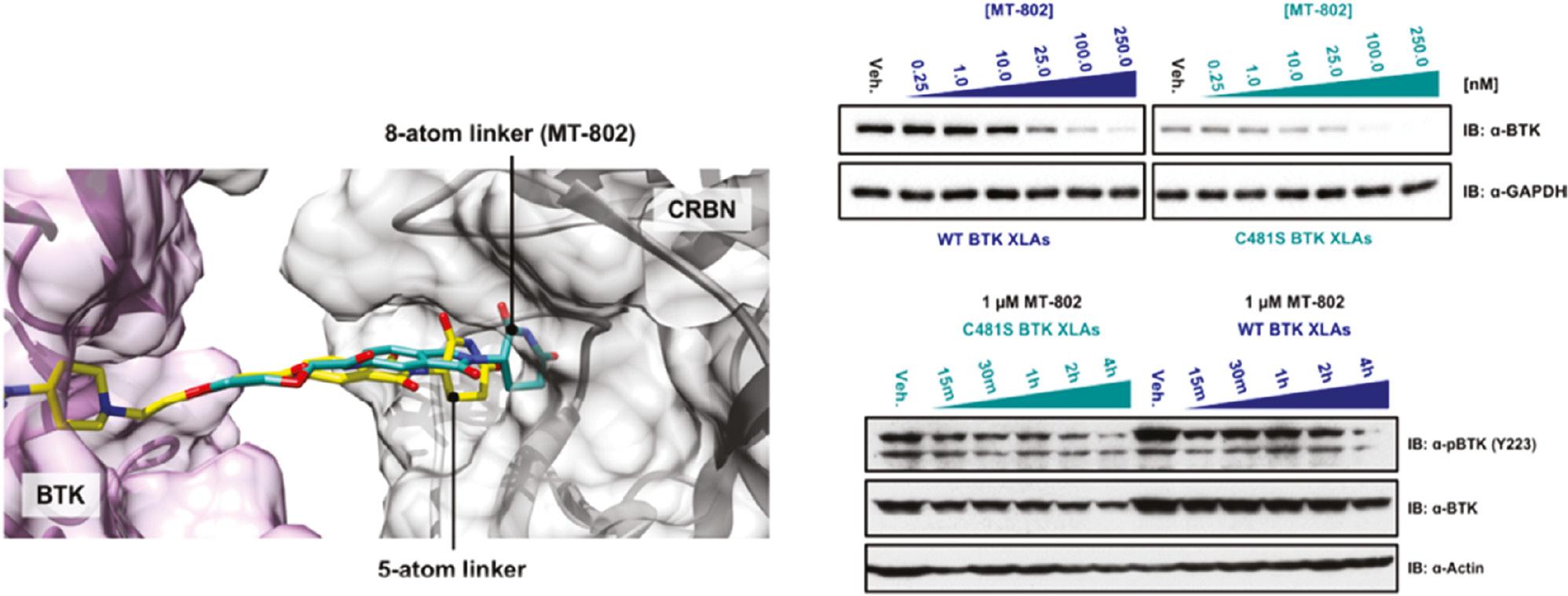
            (Left) Docking study on MT-802 (green), BTK (5P9J, purple), and cereblon (gray); (Right) MT-802 degrades wild-type and C481S-mutant BTK. Reproduced with permission from ref. [63]. Copyright 2018 American Chemical Society.

            EZH2 is the enzymatic subunit of Polycomb repressive complex 2 (PRC2), which mainly trimethylates lysine 27 of histone H3 and consequently silences gene transcription. EZH2 overexpression or gain-of-function mutations are associated with multiple cancers [65, 66]. Traditional EZH2 inhibitors focus on inhibition of methylation enzymatic activity; however, increasing evidence indicates that the protein itself is involved in tumor proliferation in a methylation-independent mechanism [6769], thus causing acquired resistance and drug insensitivity [70]. Hence, the degradation of EZH2 protein might be a potential therapeutic method for EZH2-dependent cancers. Tu et al. have reported specific PROTAC degraders of EZH2, which have shown better antitumor effects against lymphomas in vitro and in vivo than traditional EZH2 inhibitors. The authors have selected the EZH2 inhibitor EPZ6438 as the target protein ligand, and designed and synthesized two series of PROTAC-based EZH2 degraders that recruit different E3 ligase systems, von Hippel–Lindau (VHL) or CRBN. Through in vitro experiments and molecular docking model analysis ( Figure 5 ) with EZH2 (PDB ID: 5LS6) in MOE, the authors successfully identified compounds YM181 and YM281 targeting VHL E3 ligase as the best EZH2 degraders [71].

            Figure 5 |

            (Left) Docking conformation of EPZ6438 in the catalytic domain of EZH2 (PDB ID: 5 LS6) in MOE software; (Right) western blot analysis results and representative tumor images of EZH2 and H3K27me3 levels in representative SU-DHL-6 model excised tumors. Reproduced with permission from ref. [71]. Copyright 2021 American Chemical Society.

            Mutations in Src homology region 2-containing phosphatase 2 (SHP2) are associated with multiple cancers [72]. SHP2 also participates in the regulation of the immune system, as a downstream effector of the PD-1 receptor [73, 74]. Although several SHP2 small-molecule inhibitors have been reported over the past two decades, the sequence similarity between SHP1 and SHP2 has hindered their further application. Targeting SHP2 with the PROTAC strategy might offer another avenue for impeding SHP2 activity in cancer treatment [75]. Zheng and co-workers have developed a PROTAC based on SHP099, a selective allosteric inhibitor of SHP2 activity. With a docking model of the SHP099-SHP2 complex, a series of new PROTACs have been designed by using the free amino group of SHP099 to target SHP2. The molecular docking results indicated that the designed PROTACs bind SHP2 through hydrophobic interactions. In in vitro assays, the PROTAC SP4 has shown 100-times-higher inhibitory activity than SHP099 against SHP2 in HeLa cells, through suppressing the SHP2-mediated RAS/MAPK signaling pathway [76].

            Epidermal growth factor receptor (EGFR), a receptor for members of the EGF family, is a transmembrane tyrosine kinase protein involved in many human malignancies, particularly non-small-cell lung cancers (NSCLCs) [7779]. EGFR tyrosine kinase inhibitors that inhibit the activity of mutant-EGFR ATP-binding domains have been approved by the FDA for the treatment of NSCLCs. However, drug resistance due to multipoint EGFR mutations has been observed [80]. EGFR-targeting PROTACs based on pomalidomide to degrade both EGFREx19del and EGFRL858R/T790M resistant proteins have been reported [81]. Aboelez et al. used molecular docking to design new pomalidomide-based EGFR-targeting PROTACs that can degrade both wild-type and mutant EGFR. The designed compounds have been docked against the ATP-binding sites of wild-type EGFR-TK (EGFRWT, PDB:4HJO) [82] and mutant EGFR-TK (EGFRT790M, PDB: 3W2O) with MOE ( Figure 6 ) [83]. On the basis of in vitro results ( Figure 6 ), compound 16 displays higher EGFR degradation efficacy than the other six compounds, through forming two hydrogen bonds with the Arg779 and Lys721 amino acids, thereby binding EGFRWT, and two hydrogen bonds with the Ser720 and Lys745 amino acids, thereby binding EGFRT790M; these findings support the key role of molecular docking in PROTAC screening [84].

            Figure 6 |

            (Left) Docking model of TAK-285 with the active site of EGFRT790M in MOE software; (Right) apoptosis effects of compound 16 in different cell lines [84]. Copyright 2022 Journal of Enzyme Inhibition and Medicinal Chemistry.

            Castration-resistant prostate cancer (CRPC) is resistant to androgen deprivation therapy, and thus is impervious to AR-antagonist treatment [85]. Recent research has revealed that AR overexpression is a biomarker for CRPC, particularly the AR-V7 splice variant, which contains an N-terminal and DNA-binding domain (DBD) but lacks ligand-binding domains [8688]. Previous studies have indicated that small-molecule-based PROTAC with CRBN/cullin 4A neddylation degradation systems, such as the clinical phase 2 drug ARV-110 for prostate cancer (PCa), can degrade AR [89]. Liang et al. have designed and synthesized a group of phthalimide-based PROTAC compounds on the basis of the high-affinity AR agonist RU59063 [90]. Among all complexes, compound A16 showed the best AR binding affinity and AR degradation activity. To further verify the mechanism of AR degradation, the authors analyzed the docking model of compound A16 with AR protein (PDB: 2AXA) and the CRBN E3 ubiquitin ligase (PDB: 4CI3) in MOE, and found that A16 binds AR and E3 ubiquitin ligase through hydrogen bonding [91]. Bhumireddy et al. used proprietary computational algorithms and rational structure-activity-relationship optimization to select the best AR-V7 PROTAC degrader, compound 6, based on the AR DBD binder VPC-14228. This PROTAC degrader was designed by studying VPC-14228/AR-DBD/VHL ternary complex models in GLIDE (Maestro 9.3 suite) [92]. These docking models have provided guidance for designing a new PROTAC skeleton targeting AR for PCa.

            Cyclin-dependent kinase 9 (CDK9) is a serine/threonine kinase involved in the expression of Mcl-1, an important survival protein for breast cancer cell growth [93, 94]. The inhibition or degradation of CDK9 has been found to effectively block the ability of cancer cells to resist apoptosis [95]. Several CDK9 inhibitors have been reported to treat advanced malignancies; and one of these inhibitors, flavopiridol, has entered clinical trials [96]. However, owing to the high sequence conservation of CDK9 with other CDKs, these inhibitors show reversible inhibition and drug resistance. Bian et al. have reported a CDK9 selective degrader (11c) with the CDK9 inhibitor wogonin [97]. They added substituent groups at position 8 of the wogonin flavone scaffold after preliminary structure-activity-relationship studies and molecular docking analysis between the CDK9 kinase domain and wogonin. Subsequently, wogonin and the CRBN ligand pomalidomide were conjugated via different linkers, and a series of wogonin-based PROTACs were synthesized. In western blotting assays, compound 11c showed the greatest ability to degrade CDK9. This compound also decreases the level of Mcl-1, and increases the death of CDK9-overexpressing MCF-7 cells. Molecular docking in GOLD 5.1 has been used to study the binding between the wogonin-based PROTACs and CDK9, and has revealed a similar binding mode to that of wogonin itself, thus validating the use of molecular docking in the design of mechanism-based PROTACs.

            4. CONCLUSIONS AND PERSPECTIVES

            Traditional drug discovery methods are characterized by their time-consuming nature (10−15 years), high cost (400–800 million US dollars), and low success rates [98100]. The new PROTAC drug discovery platforms are no exception, and may be even more challenging to establish, because of the relatively high molecular weights (mostly >800 Da) of PROTAC compounds. In this context, rapid computer technology developments and the abundance of available structural, chemical, and biological data may enable the application of powerful virtual screening methods to lower the cost of drug discovery, including for PROTACs [101, 102]. Virtual screening methods, particularly molecular docking, are now routinely used in the drug discovery and design process to identify novel chemical scaffolds from large compound libraries, analyze drug-target binding, accelerate structure-activity-relationship analysis, and verify the prediction of adverse effects, among other applications [103].

            In the past two decades, the astonishing progress and increased interest and investment in targeted protein degradation by both academia and industry have indicated that PROTAC technology is growing into a critical and effective therapeutic modality. The PROTAC platform avoids the problems of gene knockdown/knockout off-target effects of siRNA therapeutics, as well as the poor cell permeability of antibodies. More than 50 proteins have been targeted for degradation with PROTAC technology, including EZH2, ER, BTK, and BRD4 [58]. Two heterobifunctional PROTACs, ARV-110 and ARV-471 (with undisclosed structures), targeting AR and ER, respectively, have entered phase 1 and phase 2 clinical trials (NCT03888612 and NCT04072952) for prostate and breast cancer, respectively [104, 105].

            This review summarized the recent applications of molecular docking and virtual screening for new PROTAC drug discovery. With the aid of molecular docking, PROTAC compounds have been identified against nine target proteins: Smad3, PGD2, BRD4, BTK, EZH2, SHP2, AR, CDK9, and EGFR. Molecular docking has played key roles in selecting the correct linker atom number, screening suitable target protein ligands, and characterizing the drug-protein-E3 ternary complex interactions.

            In the future, several challenges must be overcome. Most studies described herein have validated PROTAC activity with in vitro experiments; however, in vivo data are required for further development of the compounds as clinical candidates for subsequent pharmaceutical research. Moreover, the degradation activity of PROTACs is dependent on the E3 ligase, which is unequally distributed across cell types and tissues [106]. Therefore, the distribution of the PROTAC in the body must be considered. Finally, diseases such as cancer may develop PROTAC resistance via different mechanisms, such as mutation [107]. Here, molecular docking may be able to help optimize candidates that are effective against both wild-type and mutant variants of a target, as has been demonstrated in some of the studies described in this review.

            In our view, the roles of molecular docking and virtual screening for PROTAC drug discovery will only increase in the future with the continuing development of computing technologies and artificial intelligence [108]. Moreover, as target proteins as well as E3s are increasingly researched, the number of ligands that can be incorporated into the chimeric PROTAC platform will vastly expand. We believe that molecular docking and virtual screening, combined with the maturation of PROTAC development principles, will provide new directions for screening and identifying new PROTAC drugs to offer patients new treatment options beyond conventional therapeutics.

            ACKNOWLEDGEMENTS

            This work was supported by the Science and Technology Development Fund, Macau SAR (File no. 0007/2020/A1 and 0020/2022/A1), QRCM-IRG2022-011, and the University of Macau (MYRG2019-00002-ICMS, MYRG2022-00137-ICMS and MYRG2020-00017-ICMS), the National Natural Science Foundation of China (22101230), the Natural Science Basic Research Program of Shaanxi (2021JQ-089), and the Natural Science Foundation of Chongqing, China (cstc2021jcyj-msxmX0659).

            CONFLICTS OF INTEREST

            The authors declare no conflicts of interest.

            REFERENCES

            1. Sakamoto KM, Kim KB, Kumagai A, Mercurio F, Crews CM, Deshaies RJ. Protacs: Chimeric Molecules that Target Proteins to the Skp1-Cullin-F Box Complex for Ubiquitination and Degradation. Proceedings of the National Academy of Sciences of the United States of America. 2001. Vol. 98:8554–8559

            2. Li X, Song Y. Proteolysis-Targeting Chimera (PROTAC) for Targeted Protein Degradation and Cancer Therapy. Journal of Hematology Oncology. 2020. Vol. 13:50

            3. Yokoo H, Shibata N, Endo A, Ito T, Yanase Y, Murakami Y, et al.. Discovery of a Highly Potent and Selective Degrader Targeting Hematopoietic Prostaglandin D Synthase via In Silico Design. Journal of Medicinal Chemistry. 2021. Vol. 64:15868–15882

            4. Lai AC, Crews CM. Induced Protein Degradation: An Emerging Drug Discovery Paradigm. Nature Reviews Drug Discovery. 2017. Vol. 16:101–114

            5. Li JW, Zheng G, Kaye FJ, Wu L. PROTAC Therapy as a New Targeted Therapy for Lung Cancer. Molecular Therapy. 2022.

            6. Madan J, Ahuja VK, Dua K, Samajdar S, Ramchandra M, Giri S, et al.. PROTACs: Current Trends in Protein Degradation by Proteolysis-Targeting Chimeras. BioDrugs. 2022. Vol. 36:609–623

            7. Zou Y, Ma D, Wang Y. The PROTAC Technology in Drug Development. Cell Biochemistry and Function. 2019. Vol. 37:21–30

            8. He S, Dong G, Cheng J, Wu Y, Sheng C. Strategies for Designing Proteolysis Targeting Chimaeras (PROTACs). Medicinal Research Reviews. 2022. Vol. 42:1280–1342

            9. Sun D, Zhang J, Dong G, He S, Sheng C. Blocking Non-enzymatic Functions by PROTAC-Mediated Targeted Protein Degradation. Journal of Medicinal Chemistry. 2022. Vol. 65:14276–14288

            10. Guedeney N, Cornu M, Schwalen F, Kieffer C, Voisin-Chiret AS. PROTAC Technology: A New Drug Design for Chemical Biology with Many Challenges in Drug Discovery. Drug Discovery Today. 2022. Vol. 28:103395

            11. Yao T, Xiao H, Wang H, Xu X. Recent Advances in PROTACs for Drug Targeted Protein Research. International Journal of Molecular Sciences. 2022. Vol. 23:10328

            12. Wang Y, Jiang X, Feng F, Liu W, Sun H. Degradation of Proteins by PROTACs and Other Strategies. Acta Pharmaceutica Sinica. B. 2020. Vol. 10:207–238

            13. Saraswat AL, Vartak R, Hegazy R, Patel A, Patel K. Drug Delivery Challenges and Formulation Aspects of Proteolysis Targeting Chimera (PROTACs). Drug Discovery Today. 2022. Vol. 28:103387

            14. Gadd MS, Testa A, Lucas X, Chan KH, Chen W, Lamont DJ, et al.. Structural Basis of PROTAC Cooperative Recognition for Selective Protein Degradation. Nature Chemical Biology. 2017. Vol. 13:514–521

            15. Nowak RP, DeAngelo SL, Buckley D, He Z, Donovan KA, An J, et al.. Plasticity in Binding Confers Selectivity in Ligand-Induced Protein Degradation. Nature Chemical Biology. 2018. Vol. 14:706–714

            16. Testa A, Hughes SJ, Lucas X, Wright JE, Ciulli A. Structure-Based Design of a Macrocyclic PROTAC. Angewandte Chemie International Edition in English. 2020. Vol. 59:1727–1734

            17. Scior T, Bender A, Tresadern G, Medina-Franco JL, Martínez-Mayorga K, Langer T, et al.. Recognizing Pitfalls in Virtual Screening: A Critical Review. Journal of Chemical Information and Modeling. 2012. Vol. 52:867–881

            18. Dixon SL, Smondyrev AM, Rao SN. PHASE: A Novel Approach to Pharmacophore Modeling and 3D Database Searching. Chemical Biology & Drug Design. 2006. Vol. 67:370–372

            19. Yang C, Wang W, Liang JX, Li G, Vellaisamy K, Wong CY, et al.. A Rhodium(III)-Based Inhibitor of Lysine-Specific Histone Demethylase 1 as an Epigenetic Modulator in Prostate Cancer Cells. Journal of Medicinal Chemistry. 2017. Vol. 60:2597–2603

            20. Cheng SS, Qu YQ, Wu J, Yang GJ, Liu H, Wang W, et al.. Inhibition of the CDK9-Cyclin T1 Protein-Protein Interaction as a New Approach Against Triple-Negative Breast Cancer. Acta Pharmaceutica Sinica B. 2022. Vol. 12:1390–1405

            21. Cheng S, Yang GJ, Wang W, Ma DL, Leung CH. Discovery of a Tetrahydroisoquinoline-Based CDK9-Cyclin T1 Protein-Protein Interaction Inhibitor as an Anti-Proliferative and Anti-Migration Agent Against Triple-Negative Breast Cancer Cells. Genes & Diseases. 2022. Vol. 9:1674–1688

            22. Cheng S, Yang GJ, Wang W, Song YQ, Ko CN, Han Q, et al.. Identification of a Cytisine-Based EED-EZH2 Protein-Protein Interaction Inhibitor Preventing Metastasis in Triple-Negative Breast Cancer Cells. Acta Materia Medica. 2022. Vol. 1:197–211

            23. Li G, Liu H, Feng R, Kang TS, Wang W, Ko CN, et al.. A Bioactive Ligand-Conjugated Iridium (III) Metal-Based Complex as a Keap1-Nrf2 Protein-Protein Interaction Inhibitor against Acetaminophen-Induced Acute Liver Injury. Redox Biology. 2021. Vol. 48:102129

            24. Ma DL, Chan DSH, Leung CH. Molecular Docking for Virtual Screening of Natural Product Databases. Chemical Science. 2011. Vol. 2:1656–1665

            25. Kapetanovic IM. Computer-Aided Drug Discovery and Development (CADDD): In Silico-Chemico-Biological Approach. Chemico-Biological Interactions. 2008. Vol. 171:165–176

            26. Drummond ML, Williams CI. In Silico Modeling of PROTAC-Mediated Ternary Complexes: Validation and Application. Journal of Chemical Information and Modeling. 2019. Vol. 59:1634–1644

            27. Winter GE, Buckley DL, Paulk J, Roberts JM, Souza A, Dhe-Paganon S, et al.. DRUG DEVELOPMENT. Phthalimide Conjugation as a Strategy for In Vivo Target Protein Degradation. Science. 2015. Vol. 348:1376–1381

            28. Fuller JC, Burgoyne NJ, Jackson RM. Predicting Druggable Binding Sites at the Protein-Protein Interface. Drug Discovery Today. 2009. Vol. 14:155–161

            29. Li W, Zhang J, Guo L, Wang Q. Importance of Three-Body Problems and Protein-Protein Interactions in Proteolysis-Targeting Chimera Modeling: Insights from Molecular Dynamics Simulations. Journal of Chemical Information and Modeling. 2022. Vol. 62:523–532

            30. Lebraud H, Wright DJ, Johnson CN, Heightman TD. Protein Degradation by In-Cell Self-Assembly of Proteolysis Targeting Chimeras. ACS Central Science. 2016. Vol. 2:927–934

            31. Laurie AT, Jackson RM. Q-SiteFinder: An Energy-Based Method for the Prediction of Protein-Ligand Binding Sites. Bioinformatics (Oxford, England). 2005. Vol. 21:1908–1916

            32. Meireles LM, Dömling AS, Camacho CJ. ANCHOR: A Web Server and Database for Analysis of Protein-Protein Interaction Binding Pockets for Drug Discovery. Nucleic Acids Research. 2010. Vol. 38(Web Server issue):W407–W411

            33. Zorba A, Nguyen C, Xu Y, Starr J, Borzilleri K, Smith J, et al.. Delineating the Role of Cooperativity in the Design of Potent PROTACs for BTK. Proceedings of the National Academy of Sciences of the United States of America. 2018. Vol. 115:E7285–E7292

            34. Drummond ML, Henry A, Li H, Williams CI. Improved Accuracy for Modeling PROTAC-Mediated Ternary Complex Formation and Targeted Protein Degradation via New In Silico Methodologies. Journal of Chemical Information and Modeling. 2020. Vol. 60:5234–5254

            35. Salami J, Crews CM. Waste Disposal-An Attractive Strategy for Cancer Therapy. Science. 2017. Vol. 355:1163–1167

            36. Ohoka N, Shibata N, Hattori T, Naito M. Protein Knockdown Technology: Application of Ubiquitin Ligase to Cancer Therapy. Current Cancer Drug Targets. 2016. Vol. 16:136–146

            37. Montrose K, Krissansen GW. Design of a PROTAC that Antagonizes and Destroys the Cancer-Forming X-Protein of the Hepatitis B Virus. Biochemical and Biophysical Research Communications. 2014. Vol. 453:735–740

            38. Li JH, Huang XR, Zhu HJ, Oldfield M, Cooper M, Truong LD, et al.. Advanced Glycation end Products Activate Smad Signaling via TGF-Beta-Dependent and Independent Mechanisms: Implications for Diabetic Renal and Vascular Disease. FASEB J. 2004. Vol. 18:176–178

            39. Sato M, Muragaki Y, Saika S, Roberts AB, Ooshima A. Targeted Disruption of TGF-Beta1/Smad3 Signaling Protects Against Renal Tubulointerstitial Fibrosis Induced by Unilateral Ureteral Obstruction. The Journal of Clinical Investigation. 2003. Vol. 112:1486–1494

            40. Terada Y, Hanada S, Nakao A, Kuwahara M, Sasaki S, Marumo F. Gene Transfer of Smad7 using Electroporation of Adenovirus Prevents Renal Fibrosis in Post-Obstructed Kidney. Kidney International. 2002. Vol. 61 1 Suppl:S94–S98

            41. Fukuchi M, Imamura T, Chiba T, Ebisawa T, Kawabata M, Tanaka K, et al.. Ligand-Dependent Degradation of Smad3 by a Ubiquitin Ligase Complex of ROC1 and Associated Proteins. Molecular Biology of the Cell. 2001. Vol. 12:1431–143

            42. Wang X, Feng S, Fan J, Li X, Wen Q, Luo N. New Strategy for Renal Fibrosis: Targeting Smad3 Proteins for Ubiquitination and Degradation. Biochemical Pharmacology. 2016. Vol. 116:200–209

            43. Lu M, Liu T, Jiao Q, Ji J, Tao M, Liu Y, et al.. Discovery of a Keap1-Dependent Peptide PROTAC to Knockdown Tau by Ubiquitination-Proteasome Degradation Pathway. European Journal of Medicinal Chemistry. 2018. Vol. 146:251–259

            44. An S, Fu L. Small-Molecule PROTACs: An Emerging and Promising Approach for the Development of Targeted Therapy Drugs. EBioMedicine. 2018. Vol. 36:553–562

            45. Schneekloth AR, Pucheault M, Tae HS, Crews CM. Targeted Intracellular Protein Degradation Induced by a Small Molecule: En Route to Chemical Proteomics. Bioorganic & Medicinal Chemistry Letters. 2008. Vol. 18:5904–5908

            46. Morrow JD, Parsons WG 3rd, Roberts LJ 2nd. Release of Markedly Increased Quantities of Prostaglandin D2 In Vivo in Humans Following the Administration of Nicotinic Acid. Prostaglandins. 1989. Vol. 38:263–274

            47. Lewis RA, Soter NA, Diamond PT, Austen KF, Oates JA, Roberts LJ 2nd. Prostaglandin D2 Generation After Activation of Rat and Human Mast Cells with Anti-IgE. Journal of Immunology. 1982. Vol. 129:1627–1631

            48. Urade Y, Hayaishi O. Biochemical, Structural, Genetic, Physiological, and Pathophysiological Features of Lipocalin-Type Prostaglandin D Synthase. Biochimica et Biophysica Acta. 2000. Vol. 1482:259–271

            49. Takeshita E, Komaki H, Shimizu-Motohashi Y, Ishiyama A, Sasaki M, Takeda S. A Phase I Study of TAS-205 in Patients with Duchenne Muscular Dystrophy. Annals of Clinical and Translational Neurology. 2018. Vol. 5:1338–1349

            50. Komaki H, Maegaki Y, Matsumura T, Shiraishi K, Awano H, Nakamura A, et al.. Early Phase 2 Trial of TAS-205 in Patients with Duchenne Muscular Dystrophy. Annals of Clinical and Translational Neurology. 2020. Vol. 7:181–190

            51. Yokoo H, Shibata N, Naganuma M, Murakami Y, Fujii K, Ito T, et al.. Development of a Hematopoietic Prostaglandin D Synthase-Degradation Inducer. ACS Medicinal Chemistry Letters. 2021. Vol. 12:236–241

            52. Bandopadhayay P, Bergthold G, Nguyen B, Schubert S, Gholamin S, Tang Y, et al.. BET Bromodomain Inhibition of MYC-Amplified Medulloblastoma. Clinical Cancer Research. 2014. Vol. 20:912–925

            53. Klein K, Kabala PA, Grabiec AM, Gay RE, Kolling C, Lin LL, et al.. The Bromodomain Protein Inhibitor I-BET151 Suppresses Expression of inflammatory Genes and Matrix Degrading Enzymes in Rheumatoid Arthritis Synovial Fibroblasts. Annals of the Rheumatic Diseases. 2016. Vol. 75:422–429

            54. Dhalluin C, Carlson JE, Zeng L, He C, Aggarwal AK, Zhou MM. Structure and Ligand of a Histone Acetyltransferase Bromodomain. Nature. 1999. Vol. 399:491–496

            55. Faivre EJ, Wilcox D, Ha-Bui M, Hessler P, Sehgal V, Lu X, et al.. First-In-Class, Highly BDII-Selective BET Family Inhibitor ABBV-744 Displays Potent Anti-Tumor Activity in Androgen Receptor Positive Prostate Cancer Models and an Improved Tolerability Profile. Cancer Research. 2018. Vol. 78 13_Supplement:4960

            56. Hines J, Lartigue S, Dong H, Qian Y, Crews CM. MDM2-Recruiting PROTAC Offers Superior, Synergistic Antiproliferative Activity via Simultaneous Degradation of BRD4 and Stabilization of p53. Cancer Research. 2019. Vol. 79:251–262

            57. Zhou XL, Zhao F, Xu YT, Guan YY, Yu T, Zhang YZ, et al.. A Comprehensive Review of BET-Targeting PROTACs for Cancer Therapy. Bioorganic & Medicinal Chemistry. 2022. Vol. 73:117033

            58. Jiang F, Wei Q, Li H, Li H, Cui Y, Ma Y, et al.. Discovery of Novel Small Molecule Induced Selective Degradation of the Bromodomain and Extra-Terminal (BET) Bromodomain Protein BRD4 and BRD2 with Cellular Potencies. Bioorganic & Medicinal Chemistry. 2020. Vol. 28:115181

            59. Mohamed AJ, Yu L, Bäckesjö CM, Vargas L, Faryal R, Aints A, et al.. Bruton’s Tyrosine Kinase (Btk): Function, Regulation, and Transformation with Special Emphasis on the PH Domain. Immunological Reviews. 2009. Vol. 228:58–73

            60. Krysov S, Dias S, Paterson A, Mockridge CI, Potter KN, Smith KA, et al.. Surface IgM Stimulation Induces MEK1/2-Dependent MYC Expression in Chronic Lymphocytic Leukemia Cells. Blood. 2012. Vol. 119:170–179

            61. Woyach JA, Bojnik E, Ruppert AS, Stefanovski MR, Goettl VM, Smucker KA, et al.. Bruton’s Tyrosine Kinase (BTK) Function is Important to the Development and Expansion of Chronic Lymphocytic Leukemia (CLL). Blood. 2014. Vol. 123:1207–1213

            62. Woyach JA, Furman RR, Liu TM, Ozer HG, Zapatka M, Ruppert AS, et al.. Resistance Mechanisms for the Bruton’s Tyrosine Kinase Inhibitor Ibrutinib. The New England Journal of Medicine. 2014. Vol. 370:2286–2294

            63. Buhimschi AD, Armstrong HA, Toure M, Jaime-Figueroa S, Chen TL, Lehman AM, et al.. Targeting the C481S Ibrutinib-Resistance Mutation in Bruton’s Tyrosine Kinase using PROTAC-Mediated Degradation. Biochemistry. 2018. Vol. 57:3564–3575

            64. Jaime-Figueroa S, Buhimschi AD, Toure M, Hines J, Crews CM. Design, Synthesis and Biological Evaluation of Proteolysis Targeting Chimeras (PROTACs) as a BTK Degraders with Improved Pharmacokinetic Properties. Bioorganic & Medicinal Chemistry Letters. 2020. Vol. 30:126877

            65. Margueron R, Reinberg D. The Polycomb Complex PRC2 and its Mark in Life. Nature. 2011. Vol. 469:343–349

            66. Ntziachristos P, Tsirigos A, Van Vlierberghe P, Nedjic J, Trimarchi T, Flaherty MS, et al.. Genetic Inactivation of the Polycomb Repressive Complex 2 in T Cell Acute Lymphoblastic Leukemia. Nature Medicine. 2012. Vol. 18:298–301

            67. Leslie M. First EZH2 Inhibitor Approved-for Rare Sarcoma. Cancer Discovery. 2020. Vol. 10:333–334

            68. Xu K, Wu ZJ, Groner AC, He HH, Cai C, Lis RT, et al.. EZH2 Oncogenic Activity in Castration-Resistant Prostate Cancer Cells is Polycomb-Independent. Science. 2012. Vol. 338:1465–1469

            69. Kim KH, Kim W, Howard TP, Vazquez F, Tsherniak A, Wu JN, et al.. SWI/SNF-Mutant Cancers Depend on Catalytic and Non-Catalytic Activity of EZH2. Nature Medicine. 2015. Vol. 21:1491–1496

            70. Huang X, Yan J, Zhang M, Wang Y, Chen Y, Fu X, et al.. Targeting Epigenetic Crosstalk as a Therapeutic Strategy for EZH2-Aberrant Solid Tumors. Cell. 2018. Vol. 175:186–199.e19

            71. Tu Y, Sun Y, Qiao S, Luo Y, Liu P, Jiang ZX, et al.. Design, Synthesis, and Evaluation of VHL-Based EZH2 Degraders to Enhance Therapeutic Activity Against Lymphoma. Journal of Medicinal Chemistry. 2021. Vol. 64:10167–10184

            72. Liu Q, Qu J, Zhao M, Xu Q, Sun Y. Targeting SHP2 as a Promising Strategy for Cancer Immunotherapy. Pharmacological Research. 2020. Vol. 152:104595

            73. Yokosuka T, Takamatsu M, Kobayashi-Imanishi W, Hashimoto-Tane A, Azuma M, Saito T. Programmed Cell Death 1 Forms Negative Costimulatory Microclusters that Directly Inhibit T Cell Receptor Signaling by Recruiting Phosphatase SHP2. The Journal of Experimental Medicine. 2012. Vol. 209:1201–1217

            74. Chemnitz JM, Parry RV, Nichols KE, June CH, Riley JL. SHP-1 and SHP-2 Associate with Immunoreceptor Tyrosine-Based Switch Motif of Programmed Death 1 upon Primary Human T Cell Stimulation, but Only Receptor Ligation Prevents T Cell Activation. Journal of Immunology. 2004. Vol. 173:945–954

            75. Sun X, Rao Y. PROTACs as Potential Therapeutic Agents for Cancer Drug Resistance. Biochemistry. 2020. Vol. 59:240–249

            76. Zheng M, Liu Y, Wu C, Yang K, Wang Q, Zhou Y, et al.. Novel PROTACs for Degradation of SHP2 Protein. Bioorganic Chemistry. 2021. Vol. 110:104788

            77. Jang J, To C, De Clercq DJH, Park E, Ponthier CM, Shin BH, et al.. Mutant-Selective Allosteric EGFR Degraders are Effective Against a Broad Range of Drug-Resistant Mutations. Angewandte Chemie International Edition in English. 2020. Vol. 59:14481–14489

            78. Miller KD, Nogueira L, Mariotto AB, Rowland JH, Yabroff KR, Alfano CM, et al.. Cancer Treatment and Survivorship Statistics, 2019. CA: A Cancer Journal for Clinicans. 2019. Vol. 69:363–385

            79. Duma N, Santana-Davila R, Molina JR. Non-Small Cell Lung Cancer: Epidemiology, Screening, Diagnosis, and Treatment. Mayo Clinic Proceedings. 2019. Vol. 94:1623–1640

            80. Clasadonte J, Sharif A, Baroncini M, Prevot V. Gliotransmission by Prostaglandin e(2): A Prerequisite for GnRH Neuronal Function? Frontiers in Endocrinology (Lausanne). 2011. Vol. 2:91

            81. Qu X, Liu H, Song X, Sun N, Zhong H, Qiu X, et al.. Effective Degradation of EGFR(L858R+T790M) Mutant Proteins by CRBN-Based PROTACs through Both Proteosome and Autophagy/Lysosome Degradation Systems. European Journal of Medicinal Chemistry. 2021. Vol. 218:113328

            82. Park JH, Liu Y, Lemmon MA, Radhakrishnan R. Erlotinib Binds Both Inactive and Active Conformations of the EGFR Tyrosine Kinase Domain. The Biochemical Journal. 2012. Vol. 448:417–423

            83. Sogabe S, Kawakita Y, Igaki S, Iwata H, Miki H, Cary DR, et al.. Structure-Based Approach for the Discovery of Pyrrolo[3,2-d]Pyrimidine-Based EGFR T790M/L858R Mutant Inhibitors. ACS Medicinal Chemistry Letters. 2013. Vol. 4:201–205

            84. Aboelez MO, Belal A, Xiang G, Ma X. Design, Synthesis, and Molecular Docking Studies of Novel Pomalidomide-Based PROTACs as Potential Anti-Cancer Agents Targeting EGFR(WT) and EGFR(T790M). Journal of Enzyme Inhibition and Medicinal Chemistry. 2022. Vol. 37:1196–1211

            85. Scher HI, Fizazi K, Saad F, Taplin ME, Sternberg CN, Miller K, et al.. Increased Survival with Enzalutamide in Prostate Cancer after Chemotherapy. The New England Journal of Medicine. 2012. Vol. 367:1187–1197

            86. Zhu S, Zhao D, Yan L, Jiang W, Kim JS, Gu B, et al.. BMI1 Regulates Androgen Receptor in Prostate Cancer Independently of the Polycomb Repressive Complex 1. Nature Communications. 2018. Vol. 9:500

            87. Dehm SM, Tindall DJ. Alternatively Spliced Androgen Receptor Variants. Endocrine Related Cancer. 2011. Vol. 18:R183–R196

            88. Ho Y, Dehm SM. Androgen Receptor Rearrangement and Splicing Variants in Resistance to Endocrine Therapies in Prostate Cancer. Endocrinology. 2017. Vol. 158:1533–1542

            89. Takwale AD, Jo SH, Jeon YU, Kim HS, Shin CH, Lee HK, et al.. Design and Characterization of Cereblon-Mediated Androgen Receptor Proteolysis-Targeting Chimeras. European Journal of Medicinal Chemistry. 2020. Vol. 208:112769

            90. Teutsch G, Goubet F, Battmann T, Bonfils A, Bouchoux F, Cerede E, et al.. Non-Steroidal Antiandrogens: Synthesis and Biological Profile of High-Affinity Ligands for the Androgen Receptor. The Journal of Steroid Biochemistry and Molecular Biology. 1994. Vol. 48:111–119

            91. Liang JJ, Xie H, Yang RH, Wang N, Zheng ZJ, Zhou C, et al.. Designed, Synthesized and Biological Evaluation of Proteolysis Targeting Chimeras (PROTACs) as AR Degraders for Prostate Cancer Treatment. Bioorganic & Medicinal Chemistry. 2021. Vol. 45:116331

            92. Bhumireddy A, Bandaru NVMR, Raghurami Reddy B, Gore ST, Mukherjee S, Balasubramanian WR, et al.. Design, Synthesis, and Biological Evaluation of Phenyl Thiazole-Based AR-V7 Degraders. Bioorganic & Medicinal Chemistry Letters. 2022. Vol. 55:128448

            93. Cho SJ, Kim YJ, Surh YJ, Kim BM, Lee SK. Ibulocydine is a Novel Prodrug Cdk Inhibitor that Effectively Induces Apoptosis in Hepatocellular Carcinoma Cells. The Journal of Biological Chemistry. 2011. Vol. 286:19662–19671

            94. Yin T, Lallena MJ, Kreklau EL, Fales KR, Carballares S, Torrres R, et al.. A Novel CDK9 Inhibitor Shows Potent Antitumor Efficacy in Preclinical Hematologic Tumor Models. Molecular Cancer Therapeutics. 2014. Vol. 13:1442–1456

            95. Sonawane YA, Taylor MA, Napoleon JV, Rana S, Contreras JI, Natarajan A. Cyclin Dependent Kinase 9 Inhibitors for Cancer Therapy. Journal of Medicinal Chemistry. 2016. Vol. 59:8667–8684

            96. Zeidner JF, Foster MC, Blackford AL, Litzow MR, Morris LE, Strickland SA, et al.. Randomized Multicenter Phase II Study of Flavopiridol (Alvocidib), Cytarabine, and Mitoxantrone (FLAM) versus Cytarabine/Daunorubicin (7+3) in Newly Diagnosed Acute Myeloid Leukemia. Haematologica. 2015. Vol. 100:1172–1179

            97. Bian J, Ren J, Li Y, Wang J, Xu X, Feng Y, et al.. Discovery of Wogonin-Based PROTACs Against CDK9 and Capable of Achieving Antitumor Activity. Bioorganic Chemistry. 2018. Vol. 81:373–381

            98. Hammel B, Michel MC. Why are New Drugs Expensive and How Can They Stay Affordable? Handbook of Experimental Pharmacology. 2019. Vol. 260:453–466

            99. Khanna I. Drug discovery in Pharmaceutical Industry: Productivity Challenges and Trends. Drug Discovery Today. 2012. Vol. 17:1088–1102

            100. Prasad V, Mailankody S. Research and Development Spending to Bring a Single Cancer Drug to Market and Revenues after Approval. JAMA Internal Medicine. 2017. Vol. 177:1569–1575

            101. Macalino SJ, Gosu V, Hong S, Choi S. Role of Computer-Aided Drug Design in Modern Drug Discovery. Archives of Pharmacal Research. 2015. Vol. 38:1686–1701

            102. Jorgensen WL. The Many Roles of Computation in Drug Discovery. Science. 2004. Vol. 303:1813–1818

            103. Pinzi L, Rastelli G. Molecular Docking: Shifting Paradigms in Drug Discovery. International Journal of Molecular Sciences. 2019. Vol. 20:4331

            104. Snyder LB, Neklesa TK, Chen X, Dong H, Ferraro C, Gordon DA, et al.. Discovery of ARV-110, A First in Class Androgen Receptor Degrading PROTAC for the Treatment of Men with Metastatic Castration Resistant Prostate Cancer. Cancer Research. 2021. Vol. 81 13_Supplement:43

            105. Snyder LB, Flanagan JJ, Qian Y, Gough SM, Andreoli M, Bookbinder M, et al.. The Discovery of ARV-471, An Orally Bioavailable Estrogen Receptor Degrading PROTAC for the Treatment of Patients with Breast CancerProc. 112th Annual Meeting of the American Association for Cancer Research; 2021. p. 1116

            106. Lai AC, Toure M, Hellerschmied D, Salami J, Jaime-Figueroa S, Ko E, et al.. Modular PROTAC Design for the Degradation of Oncogenic BCR-ABL. Angewandte Chemie International Edition in English. 2016. Vol. 55:807–810

            107. Zhang L, Riley-Gillis B, Vijay P, Shen Y. Acquired Resistance to BET-PROTACs (Proteolysis-Targeting Chimeras) Caused by Genomic Alterations in Core Components of E3 Ligase Complexes. Molecular Cancer Therapeutics. 2019. Vol. 18:1302–1311

            108. Stone JE, Hardy DJ, Ufimtsev IS, Schulten K. GPU-Accelerated Molecular Modeling Coming of Age. Journal of Molecular Graphics & Modeling. 2010. Vol. 29:116–125

            Author and article information

            Journal
            amm
            Acta Materia Medica
            Compuscript (Ireland )
            2737-7946
            03 February 2023
            : 2
            : 1
            : 42-53
            Affiliations
            [a ]State Key Laboratory of Quality Research in Chinese Medicine, Institute of Chinese Medical Sciences, University of Macau, Taipa, Macao 999078, China
            [b ]Institute of Medical Research, Northwestern Polytechnical University, 127 West Youyi Road, Xi’an, Shaanxi 710072, China
            [c ]Department of Biomedical Sciences, Faculty of Health Sciences, University of Macau, Macao 999078, China
            Author notes
            *Correspondence: duncanleung@ 123456um.edu.mo (C-H. Leung); whwang0206@ 123456nwpu.edu.cn (W. Wang)
            Article
            10.15212/AMM-2022-0041
            511c1b9a-8c82-4045-8ef0-59cd6b9b26bd
            Copyright © 2023 The Authors.

            Creative Commons Attribution 4.0 International License

            History
            : 20 October 2022
            : 03 January 2023
            : 15 January 2023
            Page count
            Figures: 6, Tables: 2, References: 108, Pages: 12
            Categories
            Mini Review

            Toxicology,Pathology,Biochemistry,Clinical chemistry,Pharmaceutical chemistry,Pharmacology & Pharmaceutical medicine
            Degradation,molecular docking,E3 ligase,Protein-protein interaction,Drug discovery,Small-molecule,PROTAC,Virtual screening

            Comments

            Comment on this article