821
views
0
recommends
+1 Recommend
2 collections
    0
    shares
      scite_
       
      • Record: found
      • Abstract: found
      • Article: found
      Is Open Access

      In silico identification of ivermectin as an influenza A virus nuclear export protein inhibitor

      research-article
      Bookmark

            Abstract

            Influenza A virus (IAV) is an etiological agent infecting animals and humans that is responsible for seasonal epidemics and devastating pandemics. IAV nuclear export protein (NEP) is a multifaceted protein that plays a pivotal role in the virus life cycle. One of the most important functions of IAV NEP is to transport newly synthesized viral ribonucleoproteins from the nucleus to the cytoplasm. This function is achieved by the interaction between NEP and matrix protein 1 (M1) facilitated by Trp78 surrounded by negatively charged Glu residues in the M1 binding domain of NEP. In the present study, we targeted the IAV NEP with ivermectin. Utilizing in silico molecular docking, we tested ivermectin for its ability to bind NEP. We found that ivermectin strongly binds to NEP with an affinity of –7.3 kcal/mol. The ivermectin binding site identified in this study is located in the NEP-M1 protein interaction region. It is anticipated that blocking NEP-M1 protein interaction can have a considerably deleterious effect on IAV assembly and propagation. This study highlights the possibility of exploring ivermectin as a potential IAV NEP protein blocker, which could be an important therapeutic strategy in the treatment of influenza.

            Main article text

            INTRODUCTION

            Influenza viruses are enveloped viruses with a negative-sense single-stranded segmented RNA genome that belong to the Orthomyxoviridae family [1]. This family includes four genera namely influenza A virus (IAV), influenza B virus (IBV), influenza C virus (ICV), and influenza D virus (IDV) [2]. IAV infects a wide range of hosts including humans and causes seasonal epidemics and occasional pandemics in the human population, which is of great concern with respect to human health and economic well-being [3]. The genome of IAV consists of eight genomic segments.

            Nuclear export protein (NEP) (also known as non-structural protein 2, NS2) with a sequence length of 121 amino acids, is encoded by the eighth segment via alternative splicing. NEP exhibits a highly conserved amino acid sequence and plays a key role in the infectious life cycle of the influenza virus by participating in the export of viral ribonucleoprotein (vRNP) complexes from the nucleus to the cell membrane, where they are assembled into virion progeny (see Fig. 1) [4, 5]. NEP also facilitates viral budding via interaction with ATPase [6]. During the process of the transcription and translation of viral RNA, NEP regulates the accumulation of viral RNA [7].

            Fig. 1.
            The daisy chain model for NEP-mediated nuclear export of influenza virus vRNPs. Crm1 – β-importin; RanGTP – cofactor; NEP – nuclear export protein; M1 – matrix protein 1; vRNP – viral ribonucleoprotein complex; NP – nucleoprotein; PA, PB1, PB2 – viral polymerase subunits.

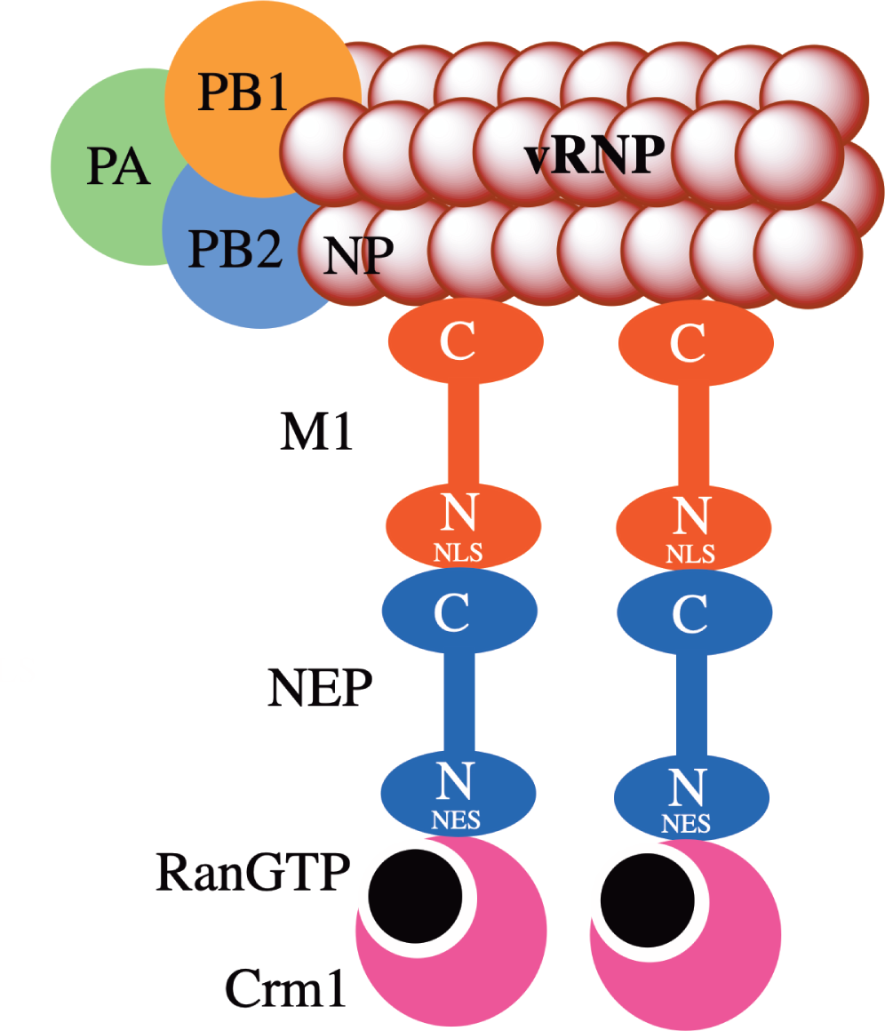
            IAV NEP comprises two domains, namely N-terminal domain (residues 1-54), which is also known as the nuclear export signal (NES) domain, and C-terminal domain (residues 63-121), which is known as M1 binding domain [8]. The N-terminal domain consists of two NESs: NES1 (residues 12-21) and NES2 (residues 31-40) [9, 10]. The C-terminal domain interacts with the nuclear localization signal (NLS) of M1 protein via exposed Trp 78 residue surrounded by a cluster of negatively charged Glu residues [11]. This interaction is critical for the export of newly synthesized viral ribonucleoproteins from the host cell nucleus to the cytoplasm [11]. The complete X-ray structure of IAV NEP has eluded researchers to date. Only the structure of the C-terminal domain of NEP has been experimentally determined [11]. The N-terminal domain of NEP adopts highly flexible and mobile confirmation that contributes to the plasticity of the NEP [12]. The C-terminal domain consists of two alpha helices that form a dimer [11].

            Ivermectin is a broad-spectrum antiparasitic drug that is widely used in veterinary medicine due to its safety and efficacy [13]. It has previously been shown that this drug has antiviral action against a broad variety of viruses. Ivermectin has been shown to act as an inhibitor of importin α/β-mediated nuclear import [14]. Ivermectin was also shown to inhibit human immunodeficiency virus (HIV-1) integrase nuclear import [14], import of the large tumor antigen of simian virus 40 [15], import of dengue virus non-structural protein 5 [14], and blocks the nuclear localization signal of parvovirus [16]. Ivermectin has been shown to impede infection caused by dengue virus 1-4 [17], West Nile Virus [18], Venezuelan equine encephalitis virus [19], influenza virus [20], pseudorabies virus [21], and severe acute respiratory syndrome coronavirus 2 [22].

            Recently, four potential binding sites of ivermectin were found on the M1 binding domain (C-terminal domain) of IAV NEP via a search for structural fragments that are similar to the structure of ivermectin binding sites of different proteins using the information from the protein data bank (PDB) [23]. In the present study, we used in silico molecular docking to test the ability of ivermectin to bind to IAV NEP. We showed that ivermectin strongly binds to IAV NEP that should block the NEP-M1 protein interaction. It is anticipated that the blocking of this interaction can have a considerably deleterious effect on IAV assembly and propagation. Therefore, ivermectin could be used as a potential IAV NEP protein blocker in the treatment of influenza that would be another successful example of drug repurposing.

            METHODS

            IAV NEP and ivermectin structure retrieval

            The protein structure of IAV NEP was retrieved from the protein data bank with the PDB identifier 1PD3 (https://www.rcsb.org/structure/1PD3) [11]. The obtained file (.pdb file) contained the structures of two chains – chain A and chain B. The chain B was removed, and the amino acid sequence 63-116 of IAV NEP that comprises chain A was analyzed. The ivermectin structure (.sdf file) was retrieved from PubChem (https://pubchem.ncbi.nlm.nih.gov/) with PubChem CID 6321424 [24].

            Molecular docking experiment

            The docking of the IAV NEP with ivermectin was carried out using the CB-Dock docking server (http://cao.labshare.cn/cb-dock/) [25]. CB-Dock is a protein-ligand docking server performing blind docking that involves four steps. In step 1, putative binding sites are detected on the protein structure. In step 2, several top binding sites are selected. The selection is based on the size of the binding sites. In step 3, the docking center is calculated and the docking box size is adjusted. In step 4, docking is carried out using AutoDock Vina and, after the completion of docking, binding sites are ranked according to the docking score [25].

            RESULTS AND DISCUSSION

            By providing an IAV NEP (.pdb file) and ivermectin structures (.sdf file) as an input to the CB-Dock server, a total of five binding sites for ivermectin were identified on NEP with varying Vina score. By default, the conformation with the highest score is treated as the best binding position and the corresponding site is considered as the optimal binding site for ivermectin [25]. Ivermectin was found to bind IAV NEP with a high AutoDock Vina score of -7.3 kcal/mol [cavity size: 185Å, center: 41*9*2 (x*y*z), and size: 29*29*29 (x*y*z)] (Fig. 2). Ivermectin interacts with the following amino acid residues at the binding site: Arg66, Glu67, Leu69, Gly70, Gln71, Glu74, Glu75, Arg77, Trp78, Leu107, Glu110, Gln111, Ile113, and Arg114. Of these, amino acids Arg66, Leu69, Glu74, Glu75, Trp78, Glu110, and Arg114 were found to be conserved [4]. Previously, four of ivermectin’s binding sites were identified on the C-terminal domain of NEP by means of the COFACTOR algorithm using the data from the protein data bank (PDB) [23]. Since these results are based on the structural similarity of ivermectin binding sites in different proteins with the structural fragments of the C-terminal domain of NEP and the authors of this study did not perform the in silico docking experiment to model the ivermectin binding to the predicted sites, these binding sites should be considered as hypothetical. According to the published data, Trp78 and the surrounding Glu residues as well as Glu81-Met100 in the M1 binding domain play a critical role in NEP-M1 protein interaction [11, 26]. The experiment described by Akarsu et al. [11] is based on the comparative study of NEP mutant proteins with one or several substitutions in amino acid sequence. Since Akarsu et al. proved that the folding of the mutant proteins corresponds to the folding of the wild type protein, their results are considered as the most reliable to date [6, 11]. The ivermectin binding site identified in this study comprises Trp78 residue as well as Glu67, Glu74, Glu75, and Glu110 residues. Since Trp78 along with the surrounding Glu residues was identified as the critical epitope for NEP binding to M1 protein [11], the NEP-M1 proteins association would be completely abolished by the interaction of IAV NEP with ivermectin because it binds to the same amino acid residues on the surface of NEP. That will prevent the export of newly synthesized viral ribonucleoproteins from the host nucleus to the cytoplasm which in turn will severely affect the life cycle of IAV. This makes ivermectin a promising anti-IAV drug molecule.

            Fig. 2.
            Docking model of ivermectin at the predicted IAV NEP site. The chemical structure of ivermectin is also shown.

            Footnotes

            Conflict of interest: The authors declare no conflicts of interest.

            Funding: The authors received no funding for this project.

            REFERENCES

            1. Wright P, Neumann G, Kawaoka Y. Fields Virology, 6th ed. Knipe DM, Howley PM, Eds.; Lippincott Williams & Wilkins: London, UK, 2013.

            2. Hause BM, Collin EA, Liu R, Huang B, Sheng Z, Lu W et al. Characterization of a novel influenza virus in cattle and Swine: proposal for a new genus in the Orthomyxoviridae family. mBio 2014;5(2), e00031-14. doi: 10.1128/mBio.00031-14.

            3. Harrington WN, Kackos CM, Webby RJ. The evolution and future of influenza pandemic preparedness. Exp Mol Med 2021; 53(5), 737-49. doi: 10.1038/s12276021-00603-0.

            4. Darapaneni V, Prabhaker VK, Kukol A. Large-scale analysis of influenza A virus sequences reveals potential drug target sites of non-structural proteins. J Gen Virol 2009; 90(9), 2124-33. doi: 10.1099/vir.0.011270-0.

            5. Li J, Yu M, Zheng W, Liu W. Nucleocytoplasmic shuttling of influenza A virus proteins. Viruses 2015; 7(5), 2668-82. doi: 10.3390/v7052668.

            6. Paterson D, Fodor E. Emerging roles for the influenza A virus nuclear export protein (NEP). PLoS Pathog 2012; 8(12), e1003019. doi: 10.1371/journal.ppat.1003019.

            7. Robb NC, Smith M, Vreede FT, Fodor E. NS2/NEP protein regulates transcription and replication of the influenza virus RNA genome. J Gen Virol 2009; 90(6), 1398-407. doi: 10.1099/vir.0.009639-0.

            8. Pinto RM, Lycett S, Gaunt E, Digard P. Accessory Gene Products of Influenza A Virus. Cold Spring Harb Perspect Med 2021; 11(12), a038380. doi: 10.1101/cshperspect.a038380.

            9. O’Neill RE, Talon J, Palese P. The influenza virus NEP (NS2 protein) mediates the nuclear export of viral ribonucleoproteins. EMBO J 1998; 17(1), 288-96. doi: 10.1093/emboj/17.1.288.

            10. Huang S, Chen J, Chen Q, Wang H, Yao Y, Chen J et al. A second CRM1-dependent nuclear export signal in the influenza A virus NS2 protein contributes to the nuclear export of viral ribonucleoproteins. J Virol 2013; 87(2), 767-78. doi: 10.1128/JVI.06519-11.

            11. Akarsu H, Burmeister WP, Petosa C, Petit I, Müller CW, Ruigrok RWH et al. Crystal structure of the M1 protein-binding domain of the influenza A virus nuclear export protein (NEP/NS2). EMBO J 2003; 22(18), 4646-55. doi: 10.1093/emboj/cdg449.

            12. Lommer BS, Luo M. Structural plasticity in influenza virus protein NS2 (NEP). J Biol Chem 2002; 277(9), 7108-17. doi: 10.1074/jbc.M109045200.

            13. Gonzalez Canga A, Sahagún Prieto AM, Liébana MJD, Martínez NF, Vega MS, García Vieitez JJ. The pharmacokinetics and interactions of ivermectin in humans–a mini-review. AAPS J 2008; 10(1), 42-6. doi: 10.1208/s12248-007-9000-9.

            14. Wagstaff KM, Sivakumaran H, Heaton SM, Harrich D, Jans DA. Ivermectin is a specific inhibitor of importin α/β-mediated nuclear import able to inhibit replication of HIV-1 and dengue virus. Biochem J 2012; 443(3), 851-6. doi: 10.1042/BJ20120150.

            15. Wagstaff KM, Rawlinson SM, Hearps AC, Jans DA. An AlphaScreen®-based assay for high-through-put screening for specific inhibitors of nuclear import. J Biomol Screen 2011; 16(2), 192-200. doi: 10.1177/1087057110390360.

            16. Nguyen KY, Kitikarn S, Robert K, Leigh O. Ivermectin blocks the nuclear location signal of parvoviruses in crayfish, Cherax quadricarinatus. Aquaculture 2014; 420-421, 288-94. doi: 10.1016/j.aquaculture.2013.11.022.

            17. Tay MY, Fraser JE, Chan WK, Moreland NJ, Rathore AP, Wang C et al. Nuclear localization of dengue virus (DENV) 1-4 non-structural protein 5; protection against all 4 DENV serotypes by the inhibitor Ivermectin. Antiviral Res 2013; 99(3), 301-6. doi: 10.1016/j.antiviral.2013.06.002.

            18. Yang SNY,Atkinson SC,Wang C,Lee A,Bogoyevitch MA, Borg NA et al. The broad spectrum antiviral ivermectin targets the host nuclear transport importin α/β1 heterodimer. Antiviral Res 2020; 177, 104760. doi: 10.1016/j.antiviral.2020.104760.

            19. Lundberg L, Pinkham C, Baer A, Amaya M, Narayanan A, Wagstaff KM et al. Nuclear import and export inhibitors alter capsid protein distribution in mammalian cells and reduce Venezuelan Equine Encephalitis Virus replication. Antiviral Res 2013; 100(3), 662-72. doi: 10.1016/j.antiviral.2013.10.004.

            20. Götz V, Magar L, Dornfeld D, Giese S, Pohlmann A, Höper D et al. Influenza A viruses escape from MxA restriction at the expense of efficient nuclear vRNP import. Sci Rep 2016; 6, 23138. doi: 10.1038/srep23138.

            21. Lv C, Liu W, Wang B, Dang R, Qiu L, Ren J et al. Iver-mectin inhibits DNA polymerase UL42 of pseudorabies virus entrance into the nucleus and proliferation of the virus in vitro and vivo. Antiviral Res 2018; 159, 55-62. doi: 10.1016/j.antiviral.2018.09.010.

            22. Caly L, Druce J, Catton M, Jans D, Wagstaff K. The FDA-approved drug ivermectin inhibits the replication of SARS-CoV-2 in vitro. Antiviral Res 2020; 178, 104787. doi: 10.1016/j.antiviral.2020.104787.

            23. Darapaneni V. Novel Ivermectin Drug Binding Sites in the C-terminal Domain of Influenza A Virus Nuclear Export Protein. Am J Curr Virol 2015; 1, 13-9. doi: 10.30493/dls.2022.330732.

            24. Kim S, Thiessen PA, Bolton EE, Chen J, Fu G, Gindulyte A et al. PubChem Substance and Compound databases. Nucleic Acids Res 2016; 44(D1), D1202-13. doi: 10.1093/nar/gkv951.

            25. Liu Y, Grimm M, Dai WT, Hou MC, Xiao ZX, Cao Y. CB-Dock: a web server for cavity detection-guided protein-ligand blind docking. Acta Pharmacol Sin 2020; 41(1), 138-44. doi: 10.1038/s41401-019-0228-6.

            26. Shimizu T, Takizawa N, Watanabe K, Nagata K, Kobayashi N. Crucial role of the influenza virus NS2 (NEP) C-terminal domain in M1 binding and nuclear export of vRNP. FEBS Lett 2011; 585(1), 41-6. doi: 10.1016/j.febslet.2010.11.017.

            Author and article information

            Journal
            MIR J
            Microbiology Independent Research Journal (MIR Journal)
            Doctrine
            2500-2236
            2022
            18 May 2022
            : 9
            : 1
            : 71-74
            Affiliations
            [-1]Department of virology and computational biochemistry, Anvek Institute of Biomolecular Research, 55-18-1, Visakhapatnam, India, 530022
            [-2]Department of computational pharmacology, Anvek Institute of Biomolecular Research, 55-18-1, Visakhapatnam, India, 530022
            Author notes
            [# ] For correspondence: Vivek Darapaneni, Department of virology and computational biochemistry, Anvek Institute of Biomolecular Research, 55-18-1, Visakhapatnam, India, 530022, e-mail: vivek110385@ 123456gmail.com
            Author information
            https://orcid.org/0000-0002-7645-2893
            Article
            10.18527/2500-2236-2022-9-1-71-74
            a59fd17e-87de-4516-ab91-42c7ba0749b7
            © 2022 Darapaneni et al.

            This is an open access article distributed under the terms of the Creative Commons Attribution-NonCommercial-ShareAlike 4.0 International Public License (CC BYNC-SA), which permits unrestricted use, distribution, and reproduction in any medium, as long as the material is not used for commercial purposes, provided that the original author and source are cited.

            History
            : 16 February 2022
            : 22 April 2022
            Categories
            HYPOTHESIS

            Immunology,Pharmaceutical chemistry,Biotechnology,Pharmacology & Pharmaceutical medicine,Infectious disease & Microbiology,Microbiology & Virology
            binding site,Influenza A virus,NEP,ivermectin,M1 protein,docking

            Comments

            Comment on this article