278
views
0
recommends
+1 Recommend
1 collections
    0
    shares
      Are you tired of sifting through news that doesn't interest you?
      Personalize your Karger newsletter today and get only the news that matters to you!

      Sign up

      • Record: found
      • Abstract: found
      • Article: found
      Is Open Access

      Bilateral Crystalline Corneal Deposits as First Clinical Manifestation of Monoclonal Gammopathy: A Case Report

      case-report

      Read this article at

      ScienceOpenPublisherPMC
          There is no author summary for this article yet. Authors can add summaries to their articles on ScienceOpen to make them more accessible to a non-specialist audience.

          Abstract

          Aims

          To report the clinical and diagnostic findings of a patient with bilateral corneal deposits caused by an underlying monoclonal gammopathy.

          Methods

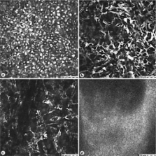
          Slit-lamp biomicroscopy, confocal microscopy and additional serological tests were performed on a 35-year-old man presenting with bilateral crystalline corneal deposits.

          Results

          The patient was diagnosed as having monoclonal gammopathy based on elevated levels of serum immunoglobulin G. Confocal microscopy showed highly reflective (protein) deposits throughout the entire cornea, with the highest density in the epithelium and anterior stromal keratocytes.

          Conclusions

          Monoclonal gammopathy, a potential sign of a life-threatening disease, can lead to dense, bilateral corneal deposits. As such changes can occur long before ocular or systemic discomforts appear, an early diagnosis is crucial. Ophthalmologists should be aware of corneal deposits as potential warning signs of monoclonal gammopathy.

          Related collections

          Most cited references6

          • Record: found
          • Abstract: found
          • Article: not found

          Immunotactoid keratopathy: a clinicopathologic case report and a review of reports of corneal involvement in systemic paraproteinemias.

          Corneal deposits in association with paraproteinemias have been well described in the ophthalmic literature. Recent reports in the renal literature have described immunotactoid deposition associated with glomerulopathy-organized microtubular deposits of IgGkappa that measured 32-50 nm in diameter on renal biopsies. We present a case of corneal immunotactoid deposition in the setting of chronic lymphocytic leukemia and review previous reports of corneal deposition in the setting of systemic paraproteinemia, highlighting the etiology, differential diagnosis, prognosis, and treatment of corneal involvement. We propose the use of the term immunotactoid keratopathy to describe corneal IgGkappa deposits appearing as tubular, electron-dense, crystalloid deposits with a central lucent core on electron microscopy and suggest that these patients undergo directed systemic workup to evaluate for potential etiologies of their systemic paraproteinemia. To illustrate the spectrum of paraprotein deposition in the cornea, and to emphasize the importance of ophthalmic evaluation in the setting of systemic paraproteinemias, we include a case of a 44-year-old man with immunoprotein corneal deposition who was subsequently diagnosed with multiple myeloma.
            Bookmark
            • Record: found
            • Abstract: not found
            • Article: not found

            Incidence of corneal crystals in the monoclonal gammopathies.

              Bookmark
              • Record: found
              • Abstract: found
              • Article: not found

              Corneal crystalline deposits associated with dysproteinemia. Report of two cases and review of the literature.

              Two patients had multiple, fine, polychromatic corneal crystals on ocular examination. Peripheral corneal biopsies and electron microscopic studies demonstrated that the crystals, which were located solely within the cytoplasm of keratocytes, exhibited an internal periodicity of 10 nm and developed within dilated cisternae of rough-surfaced endoplasmic reticulum. Histochemical studies of the corneal crystals confirmed their proteinaceous nature (immunoglobulin crystals). Serum protein analysis showed that both patients had a monoclonal gammopathy with an elevated IgG level and increased K light chains. The associated proteinopathy was unsuspected in both patients until the nature of the crystals was established. Both patients had a neoplastic process; one had a previous diagnosis of Hodgkin's disease, and multiple myeloma developed in the other on follow-up studies. Clinicians should be aware of the possibility of a malignant disorder in patients with corneal crystal-line deposits.
                Bookmark

                Author and article information

                Journal
                Case Report Ophthalmol
                COP
                Case Reports in Ophthalmology
                S. Karger AG (Allschwilerstrasse 10, P.O. Box · Postfach · Case postale, CH–4009, Basel, Switzerland · Schweiz · Suisse, Phone: +41 61 306 11 11, Fax: +41 61 306 12 34, karger@karger.ch )
                1663-2699
                May-Aug 2011
                26 July 2011
                26 July 2011
                : 2
                : 2
                : 222-227
                Affiliations
                [1] aDepartment of Ophthalmology, University Medical Center Hamburg-Eppendorf, Hamburg, Germany
                [2] bPrivate Practice, Hamburg, Germany
                Author notes
                *Dr. Johannes Steinberg, Department of Ophthalmology, University Medical Center Hamburg-Eppendorf, Martinistrasse 52, DE-20246 Hamburg (Germany), Tel. +49 40 7410 53305, E-Mail j.steinberg@ 123456uke.uni-hamburg.de
                Article
                cop0002-0222
                10.1159/000330334
                3150969
                21829405
                31aad0a2-a502-4a5b-a9c9-7c2ce8407f61
                Copyright © 2011 by S. Karger AG, Basel

                This is an Open Access article distributed under the terms of the Creative Commons Attribution-Noncommercial-No-Derivative-Works License ( http://creativecommons.org/licenses/by-nc-nd/3.0/). Users may download, print and share this work on the Internet for noncommercial purposes only, provided the original work is properly cited, and a link to the original work on http://www.karger.com and the terms of this license are included in any shared versions.

                History
                Page count
                Figures: 3, References: 7, Pages: 6
                Categories
                Published: July 2011

                Ophthalmology & Optometry
                monoclonal gammopathy,diagnosis,bilateral corneal deposits
                Ophthalmology & Optometry
                monoclonal gammopathy, diagnosis, bilateral corneal deposits

                Comments

                Comment on this article