0
views
0
recommends
+1 Recommend
0 collections
    0
    shares
      • Record: found
      • Abstract: found
      • Article: found
      Is Open Access

      Visible Light Optical Coherence Tomography Reveals the Relationship of the Myoid and Ellipsoid to Band 2 in Humans

      research-article

      Read this article at

      Bookmark
          There is no author summary for this article yet. Authors can add summaries to their articles on ScienceOpen to make them more accessible to a non-specialist audience.

          Abstract

          Purpose

          We employ visible light optical coherence tomography (OCT) to investigate the relationship between the myoid, ellipsoid, and band 2 in the living human retina. Rather than refute existing theories, we aim to reveal new bands and better delineate the structures at hand.

          Methods

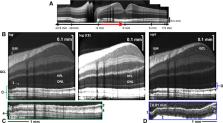
          An upgraded spectral/Fourier domain visible light OCT prototype, with 1.0-µm axial resolution, imaged 13 eyes of 13 young adult human subjects (23–40 years old) without a history of ocular pathology. The external limiting membrane (band 1) and band 2 edges were segmented. Reflectivity was examined along the inner segment (IS), defined as extending from band 1 to the band 2 center, and within band 2 itself.

          Results

          Images highlight a nearly continuously resolved extrafoveal internal limiting membrane, the peripheral single-cell thick ganglion cell layer, and the peripheral photoreceptor axonal fiber layer, a peripheral division of band 2 into bands 2a and 2b, and a reflectivity-based division of the IS into “m” and “e” zones.

          Discussion

          Topography and transverse intensity variations of the outermost band 2b suggest an association with rods. The “m” and “e” zone border is consistent with the myoid–ellipsoid boundary, even recapitulating the well-documented distribution of mitochondria throughout the IS at the foveal center. Theories of outer retinal reflectivity in OCT must adequately explain these observations.

          Translational Relevance

          Findings support that band 2 does partially overlap with the ellipsoid in transversally averaged OCT images due to photoreceptor IS length dispersion but argue that the inner ellipsoid must be inner to band 2, as suggested by prior quantitative measurements.

          Related collections

          Most cited references82

          • Record: found
          • Abstract: found
          • Article: not found

          Human photoreceptor topography.

          We have measured the spatial density of cones and rods in eight whole-mounted human retinas, obtained from seven individuals between 27 and 44 years of age, and constructed maps of photoreceptor density and between-individual variability. The average human retina contains 4.6 million cones (4.08-5.29 million). Peak foveal cone density averages 199,000 cones/mm2 and is highly variable between individuals (100,000-324,000 cones/mm2). The point of highest density may be found in an area as large as 0.032 deg2. Cone density falls steeply with increasing eccentricity and is an order of magnitude lower 1 mm away from the foveal center. Superimposed on this gradient is a streak of high cone density along the horizontal meridian. At equivalent eccentricities, cone density is 40-45% higher in nasal compared to temporal retina and slightly higher in midperipheral inferior compared to superior retina. Cone density also increases slightly in far nasal retina. The average human retina contains 92 million rods (77.9-107.3 million). In the fovea, the average horizontal diameter of the rod-free zone is 0.350 mm (1.25 degrees). Foveal rod density increases most rapidly superiorly and least rapidly nasally. The highest rod densities are located along an elliptical ring at the eccentricity of the optic disk and extending into nasal retina with the point of highest density typically in superior retina (5/6 eyes). Rod densities decrease by 15-25% where the ring crosses the horizontal meridian. Rod density declines slowly from the rod ring to the far periphery and is highest in nasal and superior retina. Individual variability in photoreceptor density differs with retinal region and is similar for both cones and rods. Variability is highest near the fovea, reaches a minimum in the midperiphery, and then increases with eccentricity to the ora serrata. The total number of foveal cones is similar for eyes with widely varying peak cone density, consistent with the idea that the variability reflects differences in the lateral migration of photoreceptors during development. Two fellow eyes had cone and rod numbers within 8% and similar but not identical photoreceptor topography.
            Bookmark
            • Record: found
            • Abstract: found
            • Article: not found

            Müller cells in the healthy and diseased retina.

            Müller glial cells span the entire thickness of the tissue, and ensheath all retinal neurons, in vertebrate retinae of all species. This morphological relationship is reflected by a multitude of functional interactions between neurons and Müller cells, including a 'metabolic symbiosis' and the processing of visual information. Müller cells are also responsible for the maintenance of the homeostasis of the retinal extracellular milieu (ions, water, neurotransmitter molecules, and pH). In vascularized retinae, Müller cells may also be involved in the control of angiogenesis, and the regulation of retinal blood flow. Virtually every disease of the retina is associated with a reactive Müller cell gliosis which, on the one hand, supports the survival of retinal neurons but, on the other hand, may accelerate the progress of neuronal degeneration: Müller cells protect neurons via a release of neurotrophic factors, the uptake and degradation of the excitotoxin, glutamate, and the secretion of the antioxidant, glutathione. However, gliotic Müller cells display a dysregulation of various neuron-supportive functions. This contributes to a disturbance of retinal glutamate metabolism and ion homeostasis, and causes the development of retinal edema and neuronal cell death. Moreover, there are diseases evoking a primary Müller cell insufficiency, such as hepatic retinopathy and certain forms of glaucoma. Any impairment of supportive functions of Müller cells, primary or secondary, must cause and/or aggravate a dysfunction and loss of neurons, by increasing the susceptibility of neurons to stressful stimuli in the diseased retina. On the contrary, Müller cells may be used in the future for novel therapeutic strategies to protect neurons against apoptosis (somatic gene therapy), or to differentiate retinal neurons from Müller/stem cells. Meanwhile, a proper understanding of the gliotic responses of Müller cells in the diseased retina, and of their protective vs. detrimental effects, is essential for the development of efficient therapeutic strategies that use and stimulate the neuron-supportive/protective-and prevent the destructive-mechanisms of gliosis.
              Bookmark
              • Record: found
              • Abstract: found
              • Article: not found

              Glaucomatous damage of the macula.

              There is a growing body of evidence that early glaucomatous damage involves the macula. The anatomical basis of this damage can be studied using frequency domain optical coherence tomography (fdOCT), by which the local thickness of the retinal nerve fiber layer (RNFL) and local retinal ganglion cell plus inner plexiform (RGC+) layer can be measured. Based upon averaged fdOCT results from healthy controls and patients, we show that: 1. For healthy controls, the average RGC+ layer thickness closely matches human histological data; 2. For glaucoma patients and suspects, the average RGC+ layer shows greater glaucomatous thinning in the inferior retina (superior visual field (VF)); and 3. The central test points of the 6° VF grid (24-2 test pattern) miss the region of greatest RGC+ thinning. Based upon fdOCT results from individual patients, we have learned that: 1. Local RGC+ loss is associated with local VF sensitivity loss as long as the displacement of RGCs from the foveal center is taken into consideration; and 2. Macular damage is typically arcuate in nature and often associated with local RNFL thinning in a narrow region of the disc, which we call the macular vulnerability zone (MVZ). According to our schematic model of macular damage, most of the inferior region of the macula projects to the MVZ, which is located largely in the inferior quadrant of the disc, a region that is particularly susceptible to glaucomatous damage. A small (cecocentral) region of the inferior macula, and all of the superior macula (inferior VF), project to the temporal quadrant, a region that is less susceptible to damage. The overall message is clear; clinicians need to be aware that glaucomatous damage to the macula is common, can occur early in the disease, and can be missed and/or underestimated with standard VF tests that use a 6° grid, such as the 24-2 VF test. Copyright © 2012 Elsevier Ltd. All rights reserved.
                Bookmark

                Author and article information

                Journal
                Transl Vis Sci Technol
                Transl Vis Sci Technol
                TVST
                Translational Vision Science & Technology
                The Association for Research in Vision and Ophthalmology
                2164-2591
                02 September 2022
                September 2022
                : 11
                : 9
                : 3
                Affiliations
                [1 ]Department of Ophthalmology, NYU Langone Health, New York, NY, USA
                [2 ]Department of Radiology, NYU Langone Health, New York, NY, USA
                [3 ]Tech4Health Institute, NYU Langone Health, New York, NY, USA
                [4 ]Department of Biomedical Engineering, University of California, Davis, CA, USA
                Author notes
                [* ] Correspondence: Vivek J. Srinivasan, Department of Radiology and Department of Ophthalmology, NYU Langone Health, NYU Langone Health, 433 1st Ave, 10th Floor, New York, NY 10010, USA. e-mail: vivek.srinivasan@ 123456nyulangone.org
                Article
                TVST-22-4522
                10.1167/tvst.11.9.3
                9440607
                36053140
                01d3738d-5fbd-4f7a-a069-15b2d970f31c
                Copyright 2022 The Authors

                This work is licensed under a Creative Commons Attribution-NonCommercial-NoDerivatives 4.0 International License.

                History
                : 03 August 2022
                : 27 March 2022
                Page count
                Pages: 14
                Categories
                Retina
                Retina

                visible light optical coherence tomography,rods,cones,inner segment/outer segment junction,ellipsoid zone

                Comments

                Comment on this article