0
views
0
recommends
+1 Recommend
0 collections
    0
    shares
      • Record: found
      • Abstract: found
      • Article: found
      Is Open Access

      The Tumor Immune Profile of Murine Ovarian Cancer Models: An Essential Tool for Ovarian Cancer Immunotherapy Research

      research-article

      Read this article at

      Bookmark
          There is no author summary for this article yet. Authors can add summaries to their articles on ScienceOpen to make them more accessible to a non-specialist audience.

          Abstract

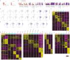
          Epithelial ovarian cancer (EOC) is the most lethal gynecologic cancer with an imperative need for new treatments. Immunotherapy has had marked success in some cancer types; however, clinical trials studying the efficacy of immune checkpoint inhibitors for the treatment of EOC benefited less than 15% of patients. Given that EOC develops from multiple tissues in the reproductive system and metastasizes widely throughout the peritoneal cavity, responses to immunotherapy are likely hindered by heterogeneous tumor microenvironments (TME) containing a variety of immune profiles. To fully characterize and compare syngeneic model systems that may reflect this diversity, we determined the immunogenicity of six ovarian tumor models in vivo, the T and myeloid profile of orthotopic tumors and the immune composition and cytokine profile of ascites, by single-cell RNA sequencing, flow cytometry, and IHC. The selected models reflect the different cellular origins of EOC (ovarian and fallopian tube epithelium) and harbor mutations relevant to human disease, including Tp53 mutation, PTEN suppression, and constitutive KRAS activation. ID8-p53 −/− and ID8-C3 tumors were most highly infiltrated by T cells, whereas STOSE and MOE-PTEN/KRAS tumors were primarily infiltrated by tumor-associated macrophages and were unique in MHC class I and II expression. MOE-PTEN/KRAS tumors were capable of forming T-cell clusters. This panel of well-defined murine EOC models reflects some of the heterogeneity found in human disease and can serve as a valuable resource for studies that aim to test immunotherapies, explore the mechanisms of immune response to therapy, and guide selection of treatments for patient populations.

          Significance:

          This study highlights the main differences in the immunogenicity and immune composition found in six different models of orthotopic ovarian cancer as an essential tool for future preclinical investigations of cancer immunotherapy.

          Related collections

          Most cited references47

          • Record: found
          • Abstract: found
          • Article: found
          Is Open Access

          Integrated analysis of multimodal single-cell data

          Summary The simultaneous measurement of multiple modalities represents an exciting frontier for single-cell genomics and necessitates computational methods that can define cellular states based on multimodal data. Here, we introduce “weighted-nearest neighbor” analysis, an unsupervised framework to learn the relative utility of each data type in each cell, enabling an integrative analysis of multiple modalities. We apply our procedure to a CITE-seq dataset of 211,000 human peripheral blood mononuclear cells (PBMCs) with panels extending to 228 antibodies to construct a multimodal reference atlas of the circulating immune system. Multimodal analysis substantially improves our ability to resolve cell states, allowing us to identify and validate previously unreported lymphoid subpopulations. Moreover, we demonstrate how to leverage this reference to rapidly map new datasets and to interpret immune responses to vaccination and coronavirus disease 2019 (COVID-19). Our approach represents a broadly applicable strategy to analyze single-cell multimodal datasets and to look beyond the transcriptome toward a unified and multimodal definition of cellular identity.
            Bookmark
            • Record: found
            • Abstract: found
            • Article: not found

            Integrated Genomic Analyses of Ovarian Carcinoma

            Summary The Cancer Genome Atlas (TCGA) project has analyzed mRNA expression, miRNA expression, promoter methylation, and DNA copy number in 489 high-grade serous ovarian adenocarcinomas (HGS-OvCa) and the DNA sequences of exons from coding genes in 316 of these tumors. These results show that HGS-OvCa is characterized by TP53 mutations in almost all tumors (96%); low prevalence but statistically recurrent somatic mutations in 9 additional genes including NF1, BRCA1, BRCA2, RB1, and CDK12; 113 significant focal DNA copy number aberrations; and promoter methylation events involving 168 genes. Analyses delineated four ovarian cancer transcriptional subtypes, three miRNA subtypes, four promoter methylation subtypes, a transcriptional signature associated with survival duration and shed new light on the impact on survival of tumors with BRCA1/2 and CCNE1 aberrations. Pathway analyses suggested that homologous recombination is defective in about half of tumors, and that Notch and FOXM1 signaling are involved in serous ovarian cancer pathophysiology.
              Bookmark
              • Record: found
              • Abstract: found
              • Article: found
              Is Open Access

              Analysis of 100,000 human cancer genomes reveals the landscape of tumor mutational burden

              Background High tumor mutational burden (TMB) is an emerging biomarker of sensitivity to immune checkpoint inhibitors and has been shown to be more significantly associated with response to PD-1 and PD-L1 blockade immunotherapy than PD-1 or PD-L1 expression, as measured by immunohistochemistry (IHC). The distribution of TMB and the subset of patients with high TMB has not been well characterized in the majority of cancer types. Methods In this study, we compare TMB measured by a targeted comprehensive genomic profiling (CGP) assay to TMB measured by exome sequencing and simulate the expected variance in TMB when sequencing less than the whole exome. We then describe the distribution of TMB across a diverse cohort of 100,000 cancer cases and test for association between somatic alterations and TMB in over 100 tumor types. Results We demonstrate that measurements of TMB from comprehensive genomic profiling are strongly reflective of measurements from whole exome sequencing and model that below 0.5 Mb the variance in measurement increases significantly. We find that a subset of patients exhibits high TMB across almost all types of cancer, including many rare tumor types, and characterize the relationship between high TMB and microsatellite instability status. We find that TMB increases significantly with age, showing a 2.4-fold difference between age 10 and age 90 years. Finally, we investigate the molecular basis of TMB and identify genes and mutations associated with TMB level. We identify a cluster of somatic mutations in the promoter of the gene PMS2, which occur in 10% of skin cancers and are highly associated with increased TMB. Conclusions These results show that a CGP assay targeting ~1.1 Mb of coding genome can accurately assess TMB compared with sequencing the whole exome. Using this method, we find that many disease types have a substantial portion of patients with high TMB who might benefit from immunotherapy. Finally, we identify novel, recurrent promoter mutations in PMS2, which may be another example of regulatory mutations contributing to tumorigenesis. Electronic supplementary material The online version of this article (doi:10.1186/s13073-017-0424-2) contains supplementary material, which is available to authorized users.
                Bookmark

                Author and article information

                Contributors
                Role: ConceptualizationRole: ResourcesRole: Data curationRole: Formal analysisRole: SupervisionRole: Funding acquisitionRole: ValidationRole: InvestigationRole: VisualizationRole: MethodologyRole: Writing - original draftRole: Writing - review and editing
                Role: ConceptualizationRole: ResourcesRole: Data curationRole: Formal analysisRole: SupervisionRole: Funding acquisitionRole: ValidationRole: InvestigationRole: VisualizationRole: MethodologyRole: Writing - original draftRole: Writing - review and editing
                Role: ResourcesRole: Data curationRole: SoftwareRole: Formal analysisRole: ValidationRole: VisualizationRole: Methodology
                Role: Data curationRole: SoftwareRole: Formal analysisRole: ValidationRole: Visualization
                Role: Methodology
                Role: Methodology
                Role: Methodology
                Role: Methodology
                Role: Writing - review and editing
                Role: SupervisionRole: Funding acquisitionRole: Writing - original draftRole: Writing - review and editing
                Journal
                Cancer Res Commun
                Cancer Res Commun
                Cancer Research Communications
                American Association for Cancer Research
                2767-9764
                June 2022
                09 June 2022
                : 2
                : 6
                : 417-433
                Affiliations
                [1 ]Cancer Therapeutics Program, Ottawa Hospital Research Institute, Ottawa, Ontario, Canada.
                [2 ]Department of Cellular and Molecular Medicine, University of Ottawa, Ottawa, Ontario, Canada.
                [3 ]Department of Pharmaceutical Sciences, College of Pharmacy, University of Illinois at Chicago, Chicago, Illinois.
                Author notes

                G.M. Rodriguez and K.J.C. Galpin contributed equally to this article.

                Corresponding Author: Barbara C. Vanderhyden, Cancer Therapeutics Program, Ottawa Hospital Research Institute, 501 Smyth Road, Ottawa, Ontario K1H 8L6, Canada. Phone: 613-737-7700, ext. 70330; E-mail: bvanderhyden@ 123456ohri.ca
                Author information
                https://orcid.org/0000-0002-5366-6047
                https://orcid.org/0000-0001-7639-6724
                https://orcid.org/0000-0001-6848-6666
                https://orcid.org/0000-0002-7271-6847
                https://orcid.org/0000-0002-7644-7189
                Article
                CRC-22-0017
                10.1158/2767-9764.CRC-22-0017
                9616009
                36311166
                05868f55-956d-4c71-9687-b81e606d4589
                © 2022 The Authors; Published by the American Association for Cancer Research

                This open access article is distributed under the Creative Commons Attribution 4.0 International (CC BY 4.0) license.

                History
                : 12 January 2022
                : 07 March 2022
                : 18 May 2022
                Page count
                Pages: 17
                Funding
                Funded by: http://dx.doi.org/10.13039/501100000024, Gouvernement du Canada | Canadian Institutes of Health Research (IRSC);
                Award ID: PJT-156139
                Award Recipient :
                Funded by: http://dx.doi.org/10.13039/100000002, HHS | National Institutes of Health (NIH);
                Award ID: CA240301
                Award Recipient :
                Funded by: http://dx.doi.org/10.13039/501100000156, FRQ | Fonds de Recherche du Québec - Santé (FRQS);
                Award Recipient :
                Funded by: http://dx.doi.org/10.13039/501100000008, Gouvernement du Canada | Health Canada (Santé Canada);
                Award Recipient :
                Funded by: http://dx.doi.org/10.13039/501100000024, Gouvernement du Canada | Canadian Institutes of Health Research (IRSC);
                Award Recipient :
                Funded by: http://dx.doi.org/10.13039/501100000024, Gouvernement du Canada | Canadian Institutes of Health Research (IRSC);
                Award Recipient :
                Categories
                Research Article
                Gynecological Cancers
                Ovarian Cancer
                Tumor Microenvironment
                Immune Cells and the Microenvironment
                Immunology
                Tumor Immunology Animal Models
                Single Cell Technologies
                Preclinical Models
                Xenograft Models
                Custom metadata
                true

                Comments

                Comment on this article