7
views
0
recommends
+1 Recommend
0 collections
    0
    shares
      • Record: found
      • Abstract: found
      • Article: found
      Is Open Access

      Efficient apoptosis requires feedback amplification of upstream apoptotic signals by effector caspase-3 or -7

      research-article

      Read this article at

      Bookmark
          There is no author summary for this article yet. Authors can add summaries to their articles on ScienceOpen to make them more accessible to a non-specialist audience.

          Abstract

          Caspase-3 and -7 are redundantly required to amplify the extrinsic and intrinsic apoptotic cascade in human leukemia.

          Abstract

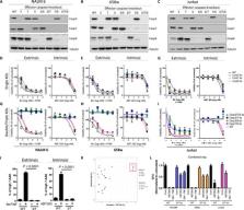
          Apoptosis is a complex multi-step process driven by caspase-dependent proteolytic cleavage cascades. Dysregulation of apoptosis promotes tumorigenesis and limits the efficacy of chemotherapy. To assess the complex interactions among caspases during apoptosis, we disrupted caspase-8, -9, -3, -7, or -6 and combinations thereof, using CRISPR-based genome editing in living human leukemia cells. While loss of apical initiator caspase-8 or -9 partially blocked extrinsic or intrinsic apoptosis, respectively, only combined loss of caspase-3 and -7 fully inhibited both apoptotic pathways, with no discernible effect of caspase-6 deficiency alone or in combination. Caspase-3/7 double knockout cells exhibited almost complete inhibition of caspase-8 or -9 activation. Furthermore, deletion of caspase-3 and -7 decreased mitochondrial depolarization and cytochrome c release upon apoptosis activation. Thus, activation of effector caspase-3 or -7 sets off explosive feedback amplification of upstream apoptotic events, which is a key feature of apoptotic signaling essential for efficient apoptotic cell death.

          Related collections

          Most cited references32

          • Record: found
          • Abstract: found
          • Article: not found

          Cleavage of BID by caspase 8 mediates the mitochondrial damage in the Fas pathway of apoptosis.

          We report here that BID, a BH3 domain-containing proapoptotic Bcl2 family member, is a specific proximal substrate of Casp8 in the Fas apoptotic signaling pathway. While full-length BID is localized in cytosol, truncated BID (tBID) translocates to mitochondria and thus transduces apoptotic signals from cytoplasmic membrane to mitochondria. tBID induces first the clustering of mitochondria around the nuclei and release of cytochrome c independent of caspase activity, and then the loss of mitochondrial membrane potential, cell shrinkage, and nuclear condensation in a caspase-dependent fashion. Coexpression of BclxL inhibits all the apoptotic changes induced by tBID. Our results indicate that BID is a mediator of mitochondrial damage induced by Casp8.
            Bookmark
            • Record: found
            • Abstract: found
            • Article: not found

            Caspases 3 and 7: key mediators of mitochondrial events of apoptosis.

            The current model of apoptosis holds that upstream signals lead to activation of downstream effector caspases. We generated mice deficient in the two effectors, caspase 3 and caspase 7, which died immediately after birth with defects in cardiac development. Fibroblasts lacking both enzymes were highly resistant to both mitochondrial and death receptor-mediated apoptosis, displayed preservation of mitochondrial membrane potential, and had defective nuclear translocation of apoptosis-inducing factor (AIF). Furthermore, the early apoptotic events of Bax translocation and cytochrome c release were also delayed. We conclude that caspases 3 and 7 are critical mediators of mitochondrial events of apoptosis.
              Bookmark
              • Record: found
              • Abstract: found
              • Article: not found

              Identification of DIABLO, a mammalian protein that promotes apoptosis by binding to and antagonizing IAP proteins.

              To identify proteins that bind mammalian IAP homolog A (MIHA, also known as XIAP), we used coimmuno-precipitation and 2D immobilized pH gradient/SDS PAGE, followed by electrospray ionization tandem mass spectrometry. DIABLO (direct IAP binding protein with low pI) is a novel protein that can bind MIHA and can also interact with MIHB and MIHC and the baculoviral IAP, OpIAP. The N-terminally processed, IAP-interacting form of DIABLO is concentrated in membrane fractions in healthy cells but released into the MIHA-containing cytosolic fractions upon UV irradiation. As transfection of cells with DIABLO was able to counter the protection afforded by MIHA against UV irradiation, DIABLO may promote apoptosis by binding to IAPs and preventing them from inhibiting caspases.
                Bookmark

                Author and article information

                Journal
                Sci Adv
                Sci Adv
                SciAdv
                advances
                Science Advances
                American Association for the Advancement of Science
                2375-2548
                July 2019
                31 July 2019
                : 5
                : 7
                : eaau9433
                Affiliations
                [1 ]Department of Oncology and Children’s Research Centre, University Children’s Hospital Zürich, 8032 Zürich, Switzerland.
                [2 ]IQVIA Technology and Services AG Theaterstrasse 4, 4051 Basel, Switzerland.
                Author notes
                [*]

                These authors contributed equally to this work.

                []Corresponding author. Email: beat.bornhauser@ 123456kispi.uzh.ch
                Author information
                http://orcid.org/0000-0002-4616-7864
                http://orcid.org/0000-0001-5290-7825
                http://orcid.org/0000-0002-9933-8356
                http://orcid.org/0000-0002-1952-0714
                http://orcid.org/0000-0003-2890-3191
                Article
                aau9433
                10.1126/sciadv.aau9433
                6669006
                31392262
                06f61e61-b78f-4c6c-8c0c-ae196fdf8fbb
                Copyright © 2019 The Authors, some rights reserved; exclusive licensee American Association for the Advancement of Science. No claim to original U.S. Government Works. Distributed under a Creative Commons Attribution NonCommercial License 4.0 (CC BY-NC).

                This is an open-access article distributed under the terms of the Creative Commons Attribution-NonCommercial license, which permits use, distribution, and reproduction in any medium, so long as the resultant use is not for commercial advantage and provided the original work is properly cited.

                History
                : 30 July 2018
                : 26 June 2019
                Funding
                Funded by: doi http://dx.doi.org/10.13039/501100011719, Institute for Empirical Research in Economics, University of Zurich;
                Funded by: Swiss Cancer League;
                Award ID: KFS 3609-02-2015 and KFS-4384-02-2018
                Funded by: Stiftung Kinderkrebsforschung Schweiz;
                Funded by: MAM-Fonds of the Children’s Research Centre of the University Children’s Hospital Zurich;
                Funded by: Fondation Panacée;
                Funded by: Novartis Foundation for biomedical research;
                Funded by: Empiris foundation,;
                Funded by: Swiss national Science Foundation SNF;
                Award ID: 310030-133108
                Funded by: Canadian institutes for health research CIHR;
                Categories
                Research Article
                Research Articles
                SciAdv r-articles
                Cell Biology
                Cell Biology
                Custom metadata
                Abigael Omana

                Comments

                Comment on this article