1
views
0
recommends
+1 Recommend
0 collections
    0
    shares
      • Record: found
      • Abstract: found
      • Article: found
      Is Open Access

      Lymphatic and Immune Cell Cross-Talk Regulates Cardiac Recovery After Experimental Myocardial Infarction

      research-article

      Read this article at

      Bookmark
          There is no author summary for this article yet. Authors can add summaries to their articles on ScienceOpen to make them more accessible to a non-specialist audience.

          Abstract

          Supplemental Digital Content is available in the text.

          Abstract

          Objective:

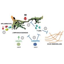
          Lymphatics play an essential pathophysiological role in promoting fluid and immune cell tissue clearance. Conversely, immune cells may influence lymphatic function and remodeling. Recently, cardiac lymphangiogenesis has been proposed as a therapeutic target to prevent heart failure after myocardial infarction (MI). We investigated the effects of gene therapy to modulate cardiac lymphangiogenesis post-MI in rodents. Second, we determined the impact of cardiac-infiltrating T cells on lymphatic remodeling in the heart.

          Approach and Results:

          Comparing adenoviral versus adeno-associated viral gene delivery in mice, we found that only sustained VEGF (vascular endothelial growth factor)-C C156S therapy, achieved by adeno-associated viral vectors, increased cardiac lymphangiogenesis, and led to reduced cardiac inflammation and dysfunction by 3 weeks post-MI. Conversely, inhibition of VEGF-C/-D signaling, through adeno-associated viral delivery of soluble VEGFR3 (vascular endothelial growth factor receptor 3), limited infarct lymphangiogenesis. Unexpectedly, this treatment improved cardiac function post-MI in both mice and rats, linked to reduced infarct thinning due to acute suppression of T-cell infiltration. Finally, using pharmacological, genetic, and antibody-mediated prevention of cardiac T-cell recruitment in mice, we discovered that both CD4 + and CD8 + T cells potently suppress, in part through interferon-γ, cardiac lymphangiogenesis post-MI.

          Conclusions:

          We show that resolution of cardiac inflammation after MI may be accelerated by therapeutic lymphangiogenesis based on adeno-associated viral gene delivery of VEGF-C C156S. Conversely, our work uncovers a major negative role of cardiac-recruited T cells on lymphatic remodeling. Our results give new insight into the interconnection between immune cells and lymphatics in orchestration of cardiac repair after injury.

          Related collections

          Most cited references42

          • Record: found
          • Abstract: found
          • Article: not found

          The inflammatory response in myocardial injury, repair, and remodelling.

          Myocardial infarction triggers an intense inflammatory response that is essential for cardiac repair, but which is also implicated in the pathogenesis of postinfarction remodelling and heart failure. Signals in the infarcted myocardium activate toll-like receptor signalling, while complement activation and generation of reactive oxygen species induce cytokine and chemokine upregulation. Leukocytes recruited to the infarcted area, remove dead cells and matrix debris by phagocytosis, while preparing the area for scar formation. Timely repression of the inflammatory response is critical for effective healing, and is followed by activation of myofibroblasts that secrete matrix proteins in the infarcted area. Members of the transforming growth factor β family are critically involved in suppression of inflammation and activation of a profibrotic programme. Translation of these concepts to the clinic requires an understanding of the pathophysiological complexity and heterogeneity of postinfarction remodelling in patients with myocardial infarction. Individuals with an overactive and prolonged postinfarction inflammatory response might exhibit left ventricular dilatation and systolic dysfunction and might benefit from targeted anti-IL-1 or anti-chemokine therapies, whereas patients with an exaggerated fibrogenic reaction can develop heart failure with preserved ejection fraction and might require inhibition of the Smad3 (mothers against decapentaplegic homolog 3) cascade. Biomarker-based approaches are needed to identify patients with distinct pathophysiologic responses and to rationally implement inflammation-modulating strategies.
            Bookmark
            • Record: found
            • Abstract: found
            • Article: found
            Is Open Access

            Development and plasticity of meningeal lymphatic vessels

            Antila et al. show that meningeal lymphatic vessels in mice develop postnatally. Interruption of VEGF-C/VEGFR3 signal transduction arrests their development. In adult mice, VEGFR3 deletion and VEGFR3 blockers, including a clinically available tyrosine kinase inhibitor, induce regression of meningeal lymphatic vessels.
              Bookmark
              • Record: found
              • Abstract: found
              • Article: not found

              Immune cells control skin lymphatic electrolyte homeostasis and blood pressure.

              The skin interstitium sequesters excess Na+ and Cl- in salt-sensitive hypertension. Mononuclear phagocyte system (MPS) cells are recruited to the skin, sense the hypertonic electrolyte accumulation in skin, and activate the tonicity-responsive enhancer-binding protein (TONEBP, also known as NFAT5) to initiate expression and secretion of VEGFC, which enhances electrolyte clearance via cutaneous lymph vessels and increases eNOS expression in blood vessels. It is unclear whether this local MPS response to osmotic stress is important to systemic blood pressure control. Herein, we show that deletion of TonEBP in mouse MPS cells prevents the VEGFC response to a high-salt diet (HSD) and increases blood pressure. Additionally, an antibody that blocks the lymph-endothelial VEGFC receptor, VEGFR3, selectively inhibited MPS-driven increases in cutaneous lymphatic capillary density, led to skin Cl- accumulation, and induced salt-sensitive hypertension. Mice overexpressing soluble VEGFR3 in epidermal keratinocytes exhibited hypoplastic cutaneous lymph capillaries and increased Na+, Cl-, and water retention in skin and salt-sensitive hypertension. Further, we found that HSD elevated skin osmolality above plasma levels. These results suggest that the skin contains a hypertonic interstitial fluid compartment in which MPS cells exert homeostatic and blood pressure-regulatory control by local organization of interstitial electrolyte clearance via TONEBP and VEGFC/VEGFR3-mediated modification of cutaneous lymphatic capillary function.
                Bookmark

                Author and article information

                Journal
                Arterioscler Thromb Vasc Biol
                Arterioscler. Thromb. Vasc. Biol
                ATV
                Arteriosclerosis, Thrombosis, and Vascular Biology
                Lippincott Williams & Wilkins
                1079-5642
                1524-4636
                14 May 2020
                July 2020
                : 40
                : 7
                : 1722-1737
                Affiliations
                [1 ]From the Normandy University, UniRouen, Inserm (Institut National de la Santé et de la Recherche Médicale) UMR1096 (EnVI Laboratory), FHU REMOD-VHF, Rouen, France (H.M., A.D., V.T., I.B., J.P.H., S.R., J.R., S.F., V.R., P.M.)
                [2 ]Normandy University, UniRouen, Inserm UMR1239 (DC2N Laboratory), Mont Saint Aignan, France (D.G., Y.A.)
                [3 ]Normandy University, UniRouen, PRIMACEN, Mont Saint Aignan, France (D.S.)
                [4 ]Wihuri Research Institute and Translational Cancer Biology Program, Research Programs Unit, Faculty of Medicine, University of Helsinki, Finland (R.K., K.A.H., K.A.)
                [5 ]Institut des Maladies Métaboliques et Cardiovasculaires (I2MC), Inserm UMR1048, Université de Toulouse III, France (N.P., M.B.)
                [6 ]Normandy University, UniRouen, Inserm (Institut National de la Santé et de la Recherche Médicale) UMR1234 (PANTHER Laboratory), Rouen, France (G.R., S.A.).
                Author notes
                Correspondence to: Ebba Brakenhielm, PhD, Inserm UMR1096, Normandy University, UniRouen, Faculty of Pharmacy and Medicine, 22 Blvd Gambetta, 76183 Rouen, France. Email ebba.brakenhielm@ 123456inserm.fr
                Article
                00018
                10.1161/ATVBAHA.120.314370
                7310303
                32404007
                07417540-4036-4195-94ea-91f5f5387b35
                © 2020 The Authors.

                Arteriosclerosis, Thrombosis, and Vascular Biology is published on behalf of the American Heart Association, Inc., by Wolters Kluwer Health, Inc. This is an open access article under the terms of the Creative Commons Attribution Non-Commercial-NoDerivs License, which permits use, distribution, and reproduction in any medium, provided that the original work is properly cited, the use is noncommercial, and no modifications or adaptations are made.

                History
                : 23 October 2019
                : 30 April 2020
                Categories
                10014
                10027
                10030
                10054
                10099
                Basic Sciences
                Custom metadata
                TRUE
                VB

                heart failure,inflammation,interferon,lymphangiogenesis,macrophages

                Comments

                Comment on this article