5
views
0
recommends
+1 Recommend
2 collections
    0
    shares
      • Record: found
      • Abstract: found
      • Article: found
      Is Open Access

      Autoantibodies in COVID‐19 correlate with antiviral humoral responses and distinct immune signatures

      research-article

      Read this article at

      Bookmark
          There is no author summary for this article yet. Authors can add summaries to their articles on ScienceOpen to make them more accessible to a non-specialist audience.

          Abstract

          Background

          Several autoimmune features occur during coronavirus disease 2019 (COVID‐19), with possible implications for disease course, immunity, and autoimmune pathology. In this study, we longitudinally screened for clinically relevant systemic autoantibodies to assess their prevalence, temporal trajectory, and association with immunity, comorbidities, and severity of COVID‐19.

          Methods

          We performed highly sensitive indirect immunofluorescence assays to detect antinuclear antibodies (ANA) and antineutrophil cytoplasmic antibodies (ANCA), along with serum proteomics and virome‐wide serological profiling in a multicentric cohort of 175 COVID‐19 patients followed up to 1 year after infection, eleven vaccinated individuals, and 41 unexposed controls.

          Results

          Compared with healthy controls, similar prevalence and patterns of ANA were present in patients during acute COVID‐19 and recovery. However, the paired analysis revealed a subgroup of patients with transient presence of certain ANA patterns during acute COVID‐19. Furthermore, patients with severe COVID‐19 exhibited a high prevalence of ANCA during acute disease. These autoantibodies were quantitatively associated with higher SARS‐CoV‐2‐specific antibody titers in COVID‐19 patients and in vaccinated individuals, thus linking autoantibody production to increased antigen‐specific humoral responses. Notably, the qualitative breadth of antibodies cross‐reactive with other coronaviruses was comparable in ANA‐positive and ANA‐negative individuals during acute COVID‐19. In autoantibody‐positive patients, multiparametric characterization demonstrated an inflammatory signature during acute COVID‐19 and alterations of the B‐cell compartment after recovery.

          Conclusion

          Highly sensitive indirect immunofluorescence assays revealed transient autoantibody production during acute SARS‐CoV‐2 infection, while the presence of autoantibodies in COVID‐19 patients correlated with increased antiviral humoral immune responses and inflammatory immune signatures.

          Abstract

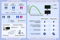
          In a multicentric cohort of 175 COVID‐19 patients, 11 vaccinated individuals, and 41 unexposed controls, we measured ANA and ANCA, along with serum proteomics and virome‐wide serological profiling. Paired analysis revealed the transient presence of ANA patterns and ANCA during acute COVID‐19. The presence of autoantibodies correlated with increased virus‐specific humoral immune responses and a proinflammatory immune signature.

          Related collections

          Most cited references58

          • Record: found
          • Abstract: found
          • Article: not found

          Clinical Characteristics of Coronavirus Disease 2019 in China

          Abstract Background Since December 2019, when coronavirus disease 2019 (Covid-19) emerged in Wuhan city and rapidly spread throughout China, data have been needed on the clinical characteristics of the affected patients. Methods We extracted data regarding 1099 patients with laboratory-confirmed Covid-19 from 552 hospitals in 30 provinces, autonomous regions, and municipalities in mainland China through January 29, 2020. The primary composite end point was admission to an intensive care unit (ICU), the use of mechanical ventilation, or death. Results The median age of the patients was 47 years; 41.9% of the patients were female. The primary composite end point occurred in 67 patients (6.1%), including 5.0% who were admitted to the ICU, 2.3% who underwent invasive mechanical ventilation, and 1.4% who died. Only 1.9% of the patients had a history of direct contact with wildlife. Among nonresidents of Wuhan, 72.3% had contact with residents of Wuhan, including 31.3% who had visited the city. The most common symptoms were fever (43.8% on admission and 88.7% during hospitalization) and cough (67.8%). Diarrhea was uncommon (3.8%). The median incubation period was 4 days (interquartile range, 2 to 7). On admission, ground-glass opacity was the most common radiologic finding on chest computed tomography (CT) (56.4%). No radiographic or CT abnormality was found in 157 of 877 patients (17.9%) with nonsevere disease and in 5 of 173 patients (2.9%) with severe disease. Lymphocytopenia was present in 83.2% of the patients on admission. Conclusions During the first 2 months of the current outbreak, Covid-19 spread rapidly throughout China and caused varying degrees of illness. Patients often presented without fever, and many did not have abnormal radiologic findings. (Funded by the National Health Commission of China and others.)
            Bookmark
            • Record: found
            • Abstract: found
            • Article: not found

            Pathophysiology, Transmission, Diagnosis, and Treatment of Coronavirus Disease 2019 (COVID-19): A Review

            The coronavirus disease 2019 (COVID-19) pandemic, due to the novel severe acute respiratory syndrome coronavirus 2 (SARS-CoV-2), has caused a worldwide sudden and substantial increase in hospitalizations for pneumonia with multiorgan disease. This review discusses current evidence regarding the pathophysiology, transmission, diagnosis, and management of COVID-19.
              Bookmark
              • Record: found
              • Abstract: found
              • Article: found
              Is Open Access

              Autoantibodies against type I IFNs in patients with life-threatening COVID-19

              The genetics underlying severe COVID-19 The immune system is complex and involves many genes, including those that encode cytokines known as interferons (IFNs). Individuals that lack specific IFNs can be more susceptible to infectious diseases. Furthermore, the autoantibody system dampens IFN response to prevent damage from pathogen-induced inflammation. Two studies now examine the likelihood that genetics affects the risk of severe coronavirus disease 2019 (COVID-19) through components of this system (see the Perspective by Beck and Aksentijevich). Q. Zhang et al. used a candidate gene approach and identified patients with severe COVID-19 who have mutations in genes involved in the regulation of type I and III IFN immunity. They found enrichment of these genes in patients and conclude that genetics may determine the clinical course of the infection. Bastard et al. identified individuals with high titers of neutralizing autoantibodies against type I IFN-α2 and IFN-ω in about 10% of patients with severe COVID-19 pneumonia. These autoantibodies were not found either in infected people who were asymptomatic or had milder phenotype or in healthy individuals. Together, these studies identify a means by which individuals at highest risk of life-threatening COVID-19 can be identified. Science, this issue p. eabd4570, p. eabd4585; see also p. 404
                Bookmark

                Author and article information

                Contributors
                onur.boyman@uzh.ch
                Journal
                Allergy
                Allergy
                10.1111/(ISSN)1398-9995
                ALL
                Allergy
                John Wiley and Sons Inc. (Hoboken )
                0105-4538
                1398-9995
                08 April 2022
                08 April 2022
                : 10.1111/all.15302
                Affiliations
                [ 1 ] Department of Immunology University Hospital Zurich Zurich Switzerland
                [ 2 ] Science for Life Laboratory Department of Women's and Children's Health Karolinska Institutet Solna Sweden
                [ 3 ] Clinic for Internal Medicine Hirslanden Klinik St. Anna Lucerne Switzerland
                [ 4 ] Department of Medicine Limmattal Hospital Schlieren Switzerland
                [ 5 ] Clinic for Internal Medicine City Hospital Triemli Zurich Zurich Switzerland
                [ 6 ] Pediatric Rheumatology Karolinska University Hospital Solna Sweden
                [ 7 ] Department of Immunology and Inflammation Imperial College London London UK
                [ 8 ] Faculty of Medicine University of Zurich Zurich Switzerland
                Author notes
                [*] [* ] Correspondence

                Onur Boyman, Department of Immunology, University Hospital Zurich, Schmelzbergstrasse 26, 8091 Zurich, Switzerland.

                Email: onur.boyman@ 123456uzh.ch

                Author information
                https://orcid.org/0000-0003-0522-7629
                https://orcid.org/0000-0001-7120-8739
                https://orcid.org/0000-0001-5387-9950
                https://orcid.org/0000-0001-7357-9090
                https://orcid.org/0000-0003-3932-788X
                https://orcid.org/0000-0002-7205-4978
                https://orcid.org/0000-0002-6161-3156
                https://orcid.org/0000-0003-2609-0246
                https://orcid.org/0000-0003-4887-7576
                https://orcid.org/0000-0001-7943-7624
                https://orcid.org/0000-0002-7896-6644
                https://orcid.org/0000-0001-5378-4716
                https://orcid.org/0000-0002-8103-0046
                https://orcid.org/0000-0001-5091-8133
                https://orcid.org/0000-0002-9046-4196
                https://orcid.org/0000-0001-8279-5545
                Article
                ALL15302
                10.1111/all.15302
                9111424
                35364615
                078945b1-db63-4c5f-b5e2-949784ff5c46
                © 2022 The Authors. Allergy published by European Academy of Allergy and Clinical Immunology and John Wiley & Sons Ltd.

                This is an open access article under the terms of the http://creativecommons.org/licenses/by-nc-nd/4.0/ License, which permits use and distribution in any medium, provided the original work is properly cited, the use is non‐commercial and no modifications or adaptations are made.

                History
                : 08 March 2022
                : 10 February 2022
                : 20 March 2022
                Page count
                Figures: 7, Tables: 2, Pages: 16, Words: 8845
                Funding
                Funded by: Universitätsspital Zürich
                Funded by: Gottfried und Julia Bangerter‐Rhyner‐Stiftung , doi 10.13039/501100005688;
                Funded by: Universität Zürich
                Funded by: Schweizerische Akademie der Medizinischen Wissenschaften , doi 10.13039/501100008485;
                Funded by: Schweizerischer Nationalfonds zur Förderung der Wissenschaftlichen Forschung
                Funded by: Digitalization Initiative of the Zurich Higher Education Institutions
                Categories
                Original Article
                ORIGINAL ARTICLES
                Autoimmunity and Clinical Immunology
                Custom metadata
                2.0
                corrected-proof
                Converter:WILEY_ML3GV2_TO_JATSPMC version:6.1.6 mode:remove_FC converted:17.05.2022

                Immunology
                antinuclear antibodies,autoantibodies,covid‐19,sars‐cov‐2,virscan
                Immunology
                antinuclear antibodies, autoantibodies, covid‐19, sars‐cov‐2, virscan

                Comments

                Comment on this article