18
views
0
recommends
+1 Recommend
1 collections
    0
    shares
      • Record: found
      • Abstract: found
      • Article: found
      Is Open Access

      Tension Pneumocephalus from Endoscopic Endonasal Surgery: A Case Series and Literature Review

      case-report

      Read this article at

      ScienceOpenPublisherPMC
      Bookmark
          There is no author summary for this article yet. Authors can add summaries to their articles on ScienceOpen to make them more accessible to a non-specialist audience.

          Abstract

          Purpose

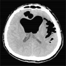
          Tension pneumocephalus (TP) caused by endoscopic endonasal surgery is a serious complication. We report four cases of TP caused by endoscopic surgery and review other cases in the literature, with special attention devoted to symptoms, imaging features, and therapeutic approaches.

          Methods

          A retrospective chart review of patients who experienced TP caused by endoscopic surgery in our institution between 2015 and 2018 was performed. Additionally, the MEDLINE database was searched for all case series or reports of TP caused by endoscopic surgery.

          Results

          Eighteen articles were identified for review, including four cases from the authors’ institution; ultimately, 26 cases were included in the present study. The main symptoms of TP were headache and a change in mental status. Cerebrospinal fluid (CSF) leakage was reported in 21 of the 26 patients (80.8%). Eight of the 26 patients (30.8%) presented with the “Mount Fuji sign” imaging feature. Twenty-four patients were treated with surgical intervention for TP (endoscopic multilayer closure of skull base defect, cranial burr hole, or bifrontal craniotomy). In addition, the present study is the first to report two patients with TP who were successfully treated conservatively.

          Conclusion

          The therapeutic method for treating TP should depend on the degree of the mass effect and clinical symptoms. When patients with TP present with obvious symptoms of CSF leakage and intracranial hypertension, urgent surgical multilayer repair of the skull base defects and/or release of the intracranial pressure are keys to treating these patients. However, conservative treatment under close observation is also feasible when the related symptoms are not overtly obvious.

          Most cited references25

          • Record: found
          • Abstract: found
          • Article: found
          Is Open Access

          Traumatic tension pneumocephalus – Two cases and comprehensive review of literature

          Although traumatic pneumocephalus is not uncommon, it rarely evolves into tension pneumocephalus (TP). Characterized by the presence of increasing amounts of intracranial air and concurrent appearance or worsening neurological symptoms, TP can be devastating if not recognized and treated promptly. We present two cases of traumatic TP and a concise review of literature on this topic. Two cases of traumatic TP are presented. In addition, a literature search revealed 20 additional cases, of which 18 had sufficient information for inclusion. Literature cases were combined with the 2 reported cases and analyzed for demographics, mechanism of injury, symptoms, time to presentation (acute 72 h), diagnostic/treatment modalities, and outcomes. Twenty cases were analyzed (17 males, 3 females, median age 26, range 8–92 years). Presentation was acute in 13/20 and delayed in 7/20 patients. Injury mechanisms included motor vehicle collisions (6/20), assault/blunt trauma to the craniofacial area (5), falls (4), and motorcycle/ bicycle crashes (3). Common presentations included depressed mental status (10/20), cerebrospinal fluid rhinorrhea (9), headache (8), and loss of consciousness (6). Computed tomography (CT) was utilized in 19/20 patients. Common underlying injuries were frontal bone/sinus fracture (9/20) and ethmoid fracture (5). Intracranial hemorrhage was seen in 5/20 patients and brain contusions in 4/20 patients. Nonoperative management was utilized in 6/20 patients. Procedural approaches included craniotomy (11/20), emergency burr hole (4), endoscopy (2), and ventriculostomy (2). Most patients responded to initial treatment (19/20). One early and one delayed death were reported. Traumatic TP is rare, tends to be associated with severe craniofacial injuries, and can occur following both blunt and penetrating injury. Early recognition and high index of clinical suspicion are important. Appropriate treatment results in improvement in vast majority of cases. CT scan is the diagnostic modality of choice for TP. Republished with permission from: Pillai P, Sharma R, MacKenzie L, Reilly EF, Beery II PR, Papadimos TJ, Stawicki SPA. Traumatic tension pneumocephalus: Two cases and comprehensive review of literature. OPUS 12 Scientist 2010;4(1):6-11.
            Bookmark
            • Record: found
            • Abstract: found
            • Article: not found

            Treatment of pneumocephalus after endoscopic sinus and microscopic skull base surgery.

            Pneumocephalus is a rare complication of endoscopic sinus surgery (ESS) and microscopic skull base surgery (MSBS). Postoperatively, patients often present with headache and altered mental status. Unrepaired leaks are associated with an increased risk of ascending meningitis. Standard treatment of pneumocephalus after ESS or MSBS has not been addressed in the literature. The study involved a retrospective review of patients at an academic tertiary care center with pneumocephalus after ESS or MSBS. Ten cases of pneumocephalus were identified, 8 after ESS and 2 after MSBS. Seven ESS defects were very small (<3 mm). The remaining three had defects more than 1 cm. Six of the 8 ESS patients had spontaneous resolution of their cerebrospinal fluid (CSF) leak and pneumocephalus, whereas all patients with larger defects failed conservative therapy with lumbar drainage. Lumbar drainage worsened the pneumocephalus in the MSBS patients. Despite resolution of pneumocephalus in many patients, all were recommended to undergo endoscopic exploration due to concern for increased risk of meningitis in unrepaired defects. Endoscopic repair was necessary in 8 cases. Repairs required a mucosal graft with or without a bone graft. Many spontaneously healed defects were found to be very tenuous. In 2 cases, patients were found to have dense scar over the defect not requiring repair. In patients with pneumocephalus after ESS or MSBS, consideration should be given to endoscopic exploration and repair of the defect with mucosal grafting. Even if spontaneous resolution has occurred, there may be increased risk of ascending meningitis through the thin or incompletely regenerated mucosa. Copyright 2010 Elsevier Inc. All rights reserved.
              Bookmark
              • Record: found
              • Abstract: found
              • Article: found
              Is Open Access

              Posttraumatic delayed subdural tension pneumocephalus

              Background: Pneumocephalus is a complication of head injury in 3.9-9.7% of the cases, it also appears after supratentorial craniotomy in 100% of cases. The accumulation of intracranial air can be acute (<72 hours) or delayed (≥72 hours). When intracranial air causes intracranial hypertension and has a mass-effect with neurological deterioration, it is called tension pneumocephalus. Case description: We represent a clinical case of a 75-year-old male patient with open penetrating head injury, complicated by tension pneumocephalus on the fifth day after trauma and underwent urgent surgical correction. Operation performed: Burr-hole placement in the right frontal region, evacuation of tension pneumocephalus. Conclusion: Tension pneumocephalus is a life-threatening neurosurgical emergency case, which needs to undergo immediate surgical or conservative treatment.
                Bookmark

                Author and article information

                Journal
                Ther Clin Risk Manag
                Ther Clin Risk Manag
                TCRM
                tcriskman
                Therapeutics and Clinical Risk Management
                Dove
                1176-6336
                1178-203X
                19 June 2020
                2020
                : 16
                : 531-538
                Affiliations
                [1 ]Department of Otolaryngology-Head and Neck Surgery, Affiliated Eye Ear Nose and Throat Hospital, Fudan University , Shanghai 200031, People’s Republic of China
                Author notes
                Correspondence: Weidong Zhao; Dehui Wang Email zhaowda@sina.com; wangdehuient@sina.com
                [*]

                These authors contributed equally to this work

                Author information
                http://orcid.org/0000-0003-1423-0942
                http://orcid.org/0000-0001-8135-7279
                http://orcid.org/0000-0003-0346-3193
                Article
                258890
                10.2147/TCRM.S258890
                7311094
                32606712
                0a1f7f78-7afb-434d-9d7c-7d9854b18872
                © 2020 Li et al.

                This work is published and licensed by Dove Medical Press Limited. The full terms of this license are available at https://www.dovepress.com/terms.php and incorporate the Creative Commons Attribution – Non Commercial (unported, v3.0) License ( http://creativecommons.org/licenses/by-nc/3.0/). By accessing the work you hereby accept the Terms. Non-commercial uses of the work are permitted without any further permission from Dove Medical Press Limited, provided the work is properly attributed. For permission for commercial use of this work, please see paragraphs 4.2 and 5 of our Terms ( https://www.dovepress.com/terms.php).

                History
                : 24 April 2020
                : 07 June 2020
                Page count
                Figures: 5, Tables: 1, References: 25, Pages: 8
                Funding
                Funded by: National Natural Science Foundation of China 10.13039/501100001809
                Award ID: 81870703
                Funded by: Joint Project of New Frontier Technology of Shanghai Shen-kang Hospital Development Center
                Award ID: SHDC 12018118
                This work was financially supported by the National Natural Science Foundation of China (No. 81870703) and the Joint Project of New Frontier Technology of Shanghai Shen-kang Hospital Development Center (SHDC 12018118).
                Categories
                Case Series

                Medicine
                tension pneumocephalus,endoscopic surgery,cerebrospinal fluid,skull base
                Medicine
                tension pneumocephalus, endoscopic surgery, cerebrospinal fluid, skull base

                Comments

                Comment on this article